226 résultats
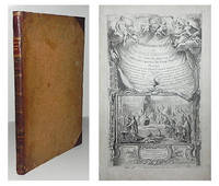
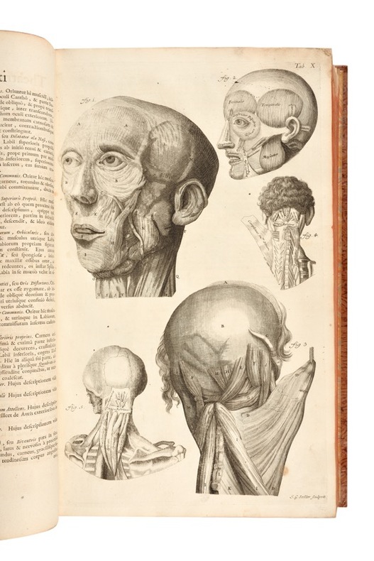
1740012020London, William Bowyer, 1740. 2 weiße Blatt, 6 Blatt, 336 S., 2 weiße Blatt. Dekorativer Lederband der Zeit mit geschmackvoll erneuertem Lederrücken. Cheseldens (1688-1752) berühmtes Lehrbuch der Anatomie erschien erstmals 1713, erlebte 13 Auflagen und wurde noch 1790 ins Deutsche übersetzt. Wie die Titelvignette veranschaulicht, wurden die Abbildungen mittels einer Camera obscura maßstabsgetreu nach medizinischen Präparaten gezeichnet und gelten als sehr korrekt. "Although Cheselden is best known for his accomplishments in the field of surgery, he wrote two important books on anatomy. The above was for many years a textbook of the English medical schools and ran through 13 editions" Garrison/Morton 390 zur Erstausgabe von 1713. Heirs of Hippocrates 812: "The fifth edition of Cheselden's noted work was printed on large and thick paper with a new series of plates engraved by Gerard van der Gucht (1696-1776) and only 250 copies were issued. Additional textual material was added and the number of plates was increased to forty with the addition of much of Cheselden's osteological work." Die Tafeln jeweils mit schwachem Druckabklatsch auf den gegenüberliegenden Textseiten. Die goldgeprägten Stehkanten gering berieben und die Vorsatzpapiere teils mit passendem, altem Marmorpapier ergänzt, sonst handelt es sich um ein ungewöhnlich dekoratives und weitgehend fleckenfreies Exemplar der schönsten Auflage des Klassikers. - One of 250 copies of the 5th edition that have been printed on thick paper and newly illustrated with Vanderguchts 40 plates. The plates each have a faint imprint on the opposite text pages. The gilt edges of the boards are slightly rubbed and some of the endpapers have been supplemented with matching old marbled paper, otherwise this is - though professionally rebacked - an unusually decorative and largely stain-free copy of the most beautiful edition of this classical work on anatomy.
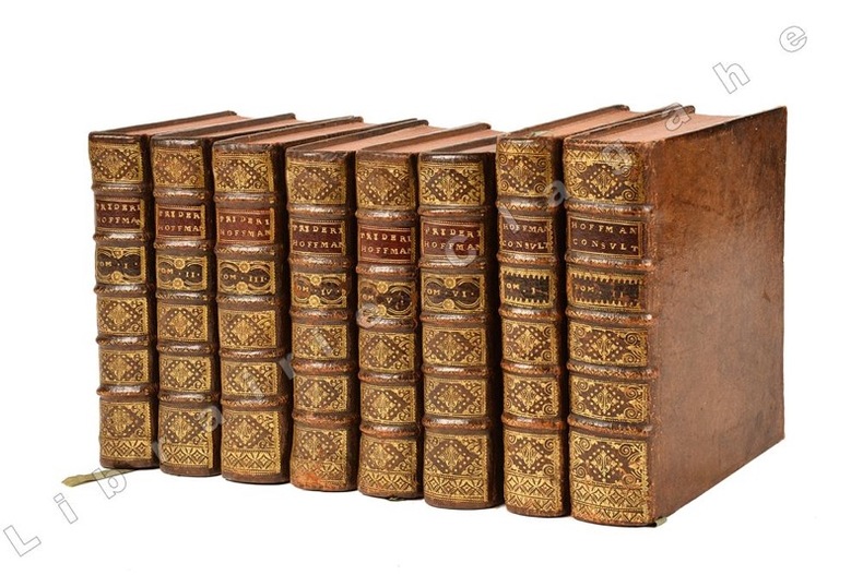
173021397Venetiis apud Ballionem, 1730-1735 ; 4 tomes reliés en 6 volumes in-4° ; portrait gravé de Hoffman par J. Rupert aux tome I, IV (1) et tome I de “Consultationum” ; le tome 4 comprend trois parties soit les tomes IV, V et VI . [Suivi de] - Consultationum et responsorum medicinalium centuria prima complectens Morbos capitis et pectoris. 1 vol., 472 pp. - Consultatiomum et responsorum medicinalium centuria secunda et tertia complectens Morbos addominis et arturum externorum. 1 vol., 864, [54] pp. Francofurti ad Moenum, Franciscum Varremtrapp, 1734-1735; soit en tout 8 volumes in-4°, veau brun marbré, dos à nerfs très décorés et dorés, pièce de titre grenat, roulette dorée sur les coupes, tranches rouges ; les 2 derniers volumes sont légèrement plus grand et le décor du dos très légèrement différent.
174830143DBGöttingen, Abraham Vandenhoeck, 1748. 4°. (4) Bl., VI S., 136 S., (1) Bl. Mit 3 gestochenen, gefalteten Tafeln. Halbleinwandband um 1850 mit goldgeprägtem Rückentitel und Rückenvergoldung. + Wichtig: Für unsere Kunden in der EU erfolgt der Versand alle 14 Tage verzollt ab Deutschland / Postbank-Konto in Deutschland vorhanden +, A|B [2 Warenabbildungen]
174325189Leiden: Joannem & Hermannum Verbeek 1743. From the first edition of one of the greatest of all Anatomical Atlases. Tabula IX which features Albinus and Wandelaar's famous "Muscle Man" a skinned figure whose musculature is visible and defined as viewed in walking profile with the left arm raised and the right foot forward his head is turned slightly away from the viewer. Elephant folio 620 by 475 mm single folio sheet now mounted with the use of none-evasive corner tabs and protected by mylar. Very well preserved fully intact with only the most minor evidence of age. ONE OF THE MAGNIFICENT PLATES FROM "AMONG THE MOST ARTISTICALLY PERFECT OF ANATOMICAL ATLASES." Wandelaar placed his skeletons and musclemen against lush ornamental backgrounds to give them the illusion of vitality using contrasts of mass and light to produce a three-dimensional effect. The most famous plate in the atlas depicts a skeletal figure standing in front of an enormous grazing rhinoceros sketched by Wandelaar from the first living specimen in Europe which had arrived at Amsterdam zoo in 1741" Norman.<br> The plates in this large folio work and in the four supplementary works in large folio with which it is bound are unsurpassed for their cool elegant aesthetic and scientific accuracy. They were drawn and engraved by Jan Wandelaer a pupil of the engravers Jacob Fokema and Guillem van der Gouwen and the painter Gerard de Lairesse who prepared the drawings for Bidloo's atlas. Prior to working for Albinus Wandelaer worked for Friedrik Ruysch. Albinus however provided Wandelaar with the opportunity for the full expression of his talents as a draftsman and engraver. <br> In an attempt to increase the scientific accuracy of anatomical illustration Albinus and Wandelaar devised a new technique of placing nets with square webbing at specified intervals between the artist and the anatomical specimen and copying the images using the grid patterns. Wandelaer placed each figure in a carefully chosen landscape setting and the artistic results are so pleasantly successful that the anatomical figures although composed of many separate parts appear to be actually stepping out of the picture. Joannem & Hermannum Verbeek unknown
174025188Leiden: Joannem & Hermannum Verbeek 1740. From the first edition of one of the greatest of all Anatomical Atlases. Tabula V which features Albinus and Wandelaar's famous "Muscle Man" a skinned figure whose musculature is visible and defined as viewed from behind standing with the left arm raised right arm turned and his weight shifted to the right foot. Elephant folio 620 by 475 mm single folio sheet now mounted with the use of none-evasive corner tabs and protected by mylar. Very well preserved fully intact with only the most minor evidence of age. ONE OF THE MAGNIFICENT PLATES FROM "AMONG THE MOST ARTISTICALLY PERFECT OF ANATOMICAL ATLASES." Wandelaar placed his skeletons and musclemen against lush ornamental backgrounds to give them the illusion of vitality using contrasts of mass and light to produce a three-dimensional effect. The most famous plate in the atlas depicts a skeletal figure standing in front of an enormous grazing rhinoceros sketched by Wandelaar from the first living specimen in Europe which had arrived at Amsterdam zoo in 1741" Norman.<br> The plates in this large folio work and in the four supplementary works in large folio with which it is bound are unsurpassed for their cool elegant aesthetic and scientific accuracy. They were drawn and engraved by Jan Wandelaer a pupil of the engravers Jacob Fokema and Guillem van der Gouwen and the painter Gerard de Lairesse who prepared the drawings for Bidloo's atlas. Prior to working for Albinus Wandelaer worked for Friedrik Ruysch. Albinus however provided Wandelaar with the opportunity for the full expression of his talents as a draftsman and engraver. <br> In an attempt to increase the scientific accuracy of anatomical illustration Albinus and Wandelaar devised a new technique of placing nets with square webbing at specified intervals between the artist and the anatomical specimen and copying the images using the grid patterns. Wandelaer placed each figure in a carefully chosen landscape setting and the artistic results are so pleasantly successful that the anatomical figures although composed of many separate parts appear to be actually stepping out of the picture. Joannem & Hermannum Verbeek unknown
174025187Leiden: Joannem & Hermannum Verbeek 1740. From the first edition of one of the greatest of all Anatomical Atlases. Tabula V which features a human skeleton in a three-quarter view and seen from the rear is portrayed in a walking motion with the left hand raised and extended. Elephant folio ca. 620 by 475 mm single folio sheet now mounted with the use of non-evasive corner tabs and protected by mylar. Very well preserved fully intact with only the most minor evidence of age. ONE OF THE MAGNIFICENT PLATES FROM "AMONG THE MOST ARTISTICALLY PERFECT OF ANATOMICAL ATLASES." Wandelaar placed his skeletons and musclemen against lush ornamental backgrounds to give them the illusion of vitality using contrasts of mass and light to produce a three-dimensional effect. The most famous plate in the atlas depicts a skeletal figure standing in front of an enormous grazing rhinoceros sketched by Wandelaar from the first living specimen in Europe which had arrived at Amsterdam Zoo in 1741" Norman.<br> The plates in this large folio work and in the four supplementary works in large folio with which it is bound are unsurpassed for their cool elegant aesthetic and scientific accuracy. They were drawn and engraved by Jan Wandelaer a pupil of the engravers Jacob Fokema and Guillem van der Gouwen and the painter Gerard de Lairesse who prepared the drawings for Bidloo's atlas. Prior to working for Albinus Wandelaer worked for Friedrik Ruysch. Albinus however provided Wandelaar with the opportunity for the full expression of his talents as a draftsman and engraver. <br> In an attempt to increase the scientific accuracy of anatomical illustration Albinus and Wandelaar devised a new technique of placing nets with square webbing at specified intervals between the artist and the anatomical specimen and copying the images using the grid patterns. Wandelaer placed each figure in a carefully chosen landscape setting and the artistic results are so pleasantly successful that the anatomical figures although composed of many separate parts appear to be actually stepping out of the picture. Joannem & Hermannum Verbeek unknown
17969532Belitz u. Braun, Berlin 1796. IV, 176 u. II, 192 S. mit 5 handkolorierten gestochenen Tafeln nach P. Mügge von C.C. Glaßbach. 4°. Interims-Broschur der Zeit. Umschläge etwas beschädigt u. fleckig. Schnitt u. im weißen Rand angestaubt. Teils etwas gebräunt u. stockfleckig. Titel mehrfach gestempelt. Unbeschnitten u. breitrandig mit sehr schönen Tafeln (Harn-, Nieren- u. Gallensteine u.a.). - Gutes Expl. Selten.
1784000285Etienne Charpentier 1784. Hardcover. See Description. Tall folio 385 mm. The entire book including the text was printed from engraved copper plates 60 plates an illustrated title page plate 29 anatomical plates and 30 calligraphic letterpress plates. Bound in a contemporary or near contemporary binding with calf spine and decorative paste paper boards - rubbed edges shelf worn old ink stain on rear cover. Both the upper portion of the spine and the tail cap are carefully restored. Board corners are neatly repaired. Interior contains an occasional minor spots of marginal foxing; a couple of tiny holes or paper defects are present on the text plate opposite plate 20. The leaves are otherwise generally clean. Francois-Michel Disdier 1708-1781 was a professor of surgery and a drawing master at the Academy of Painting in Paris. His "Tableaux anatomiques" a deluxe anatomy book for physicians was first published in 1758. Many of the plates were done by Crepy and Charpentier some after the style of Vesalius and others following Eustachi. The engraved title page designed by François Bouchet depicts students in an anatomy class studying a cadaver. The whole scene is framed in ornate baroque style. Blake NLM p. 122; Heirs of Hippocrates 894 listing 1778 edition; Hirsch II p. 190. <br/> <br/> Etienne Charpentier hardcover
1758839Paris, E. Charpentier, 1758. 60 num. gestochene Blätter (incl. Titel, 29 anatomischen Tafeln und je 1 Bl. Erklärung). Folio. Mod. Pappband mit Rückenschild.
178330052AB6 Hefte in 1 Band + Atlasband. Berlin und Leipzig, G.J. Decker und Heinrich Rottmann (Heft 5 und 6). 1783-1794. 4° bzw. Folio. Zusammen ca. 340 Textseiten. Mit 66 (6 doppelblattgross) gestochenen Tafeln. Halblederbände der Zeit mit goldgeprägtem Rückenschild und floraler Rückenvergoldung.
178330052ABBerlin und Leipzig, G.J. Decker und Heinrich Rottmann (Heft 5 und 6)., 1783-1794. 4° bzw. Folio. Zusammen ca. 340 Textseiten. Mit 66 (6 doppelblattgross) gestochenen Tafeln. Halblederbände der Zeit mit goldgeprägtem Rückenschild und floraler Rückenvergoldung. 6 Hefte in 1 Band + Atlasband.
176130077DBLeiden, Johann und Hermann Verbeek, 1761. Folio. (2) Bl., 295, 1 S. Mit 89 Kupfertafeln. Halblederband aus der Zeit mit goldgeprägtem Rückenschild.
176130077DBLeiden, Johann und Hermann Verbeek, 1761. Folio. (2) Bl., 295, 1 S. Mit 89 Kupfertafeln. Halblederband aus der Zeit mit goldgeprägtem Rückenschild. + Wichtig: Für unsere Kunden in der EU erfolgt der Versand alle 14 Tage verzollt ab Deutschland / Postbank-Konto in Deutschland vorhanden +, A
173747215ABLeyden, Johannes van Kerckhem 1737-38. 2°. [24] Bl., 452 (recte 450) S., [11] Bl. Halbleder der Zeit auf 6 Bünden mit Rückenschild und etwas Rückenvergoldung. Etwas berieben, ein Gelenk geringf. angeplatzt. Deckel-Ecken bestoßen. Eine Tafel mit kl. Wasserrand, sonst ganz sauber.
175840192ABNürnberg, bey Johann Josef Fleischmann, 1758. Gross-Folio. Titel, 41 S. Mit 39 gestochenen Tafeln. Etwas späterer Halblederband mit goldgeprägtem Rückenschild und wenig Rückenvergoldung.
175840192ABNürnberg, bey Johann Josef Fleischmann, 1758. Gross-Folio. Titel, 41 S. Mit 39 gestochenen Tafeln. Etwas späterer Halblederband mit goldgeprägtem Rückenschild und wenig Rückenvergoldung. [2 Warenabbildungen]
171610857DBGenevae (Genf), Cramer & Perachon, 1716. Folio. 42 cm. Vortitel in Schwarz, Titelblatt in Rot und Schwarz, [8] Blatt, 434 Seiten, Titelblatt in Schwarz [1] Blatt, 452 Seiten, Titelblatt in Rot und Schwarz, [5] Blatt, XVI, 34 Seiten, [5] Blatt (Index). Ganzlederbände der Zeit mit goldgeprägtem Rückentitel auf roten Lederschildchen, floraler goldgeprägter Buchschmuck. [11 Warenabbildungen] Jean-Jacques Manget, (latinisiert Johann Jacob Mangetus, geboren am 19. Juni 1652 in Genf; gestorben am 15. August 1742
171721598Genevae, Cramer et Perachon, 1717 ; 2 tomes in-folio (428 mm), demi-veau fauve du temps, dos à nerfs décoré de filets dorés, titre doré, pièce de tomaison foncée (plats refaits, gardes renouvelées) ; [20], 434 pp., [1 bl.] f. ; [4], 452 ; [12], XVI, 34, [10] pp. et en tout: portrait de Jean Jacques Manget gravé par Seiller, d’après B. Guillibaud, en frontispice, grande vignette répétée aux 3 titres, figurant une scène de dissection, gravée par Seiller et 136 planches anatomiques hors-texte ( 110 + 5 extra-ordinaires + 21 Eustache) gravées par Seiller, Gottfried Pfauntz, J.d. Hartman, Andréas Geyer et Augusta Vind.
171745024AB3 in 2 Bänden 1 Atlasband. Genève, Cramer & Perachon, 1717. Folio. 10 n.n. Bl., 434 S.; 2 n.n. Bl., 452 S., 6 n.n. Bl., XVI, 34 S., 5 n.n. Bl. (Index). Mit 3 gestochenen Titelvignetten und 136 Kupfertafeln (davon 1 mehrfach gefaltet). Halblederbände.
171735024AB3 in 2 Bänden + 1 Atlasband. Genève, Cramer & Perachon, 1717. Folio. (10) Bl., 434 S.; (2) Bl., 452 S., (6) Bl., XVI, 34 S., (5) Bl. (Index). Mit 3 gestochenen Titelvignetten und 136 Kupfertafeln (davon 1 mehrfach gefaltet). Halblederbände aus der Zeit.
171745024ABGenève, Cramer & Perachon, 1717. Folio. 10 n.n. Bl., 434 S.; 2 n.n. Bl., 452 S., 6 n.n. Bl., XVI, 34 S., 5 n.n. Bl. (Index). Mit 3 gestochenen Titelvignetten und 136 Kupfertafeln (davon 1 mehrfach gefaltet). Halblederbände. 3 in 2 Bänden 1 Atlasband.
172260290ABAmsterdam, R. & G.Wetstenisu, 1722. 36x23 cm. XLIV, 115 S. (1) w. S., (11) S. Index, (1) w. S. Mit einer gestoch. Titelvignette und 47 Kupfertafeln (davon 6 Tafeln in Altkolorit). Ldr.-Bd. der Zeit mit Rückenschild und Rückenvergoldung.
171735024ABGenève, Cramer & Perachon, 1717. Folio. (10) Bl., 434 S.; (2) Bl., 452 S., (6) Bl., XVI, 34 S., (5) Bl. (Index). Mit 3 gestochenen Titelvignetten und 136 Kupfertafeln (davon 1 mehrfach gefaltet). Halblederbände aus der Zeit. + Wichtig: Für unsere Kunden in der EU erfolgt der Versand alle 14 Tage verzollt ab Deutschland / Postbank-Konto in Deutschland vorhanden +, A 3 in 2 Bänden + 1 Atlasband.
1739ST12883Leyden: Johann Arnold Langerak 1739. First Edition in Latin. 540 x 380 mm. 21 1/4 x 15". 70 leaves of text.Translated by William Dundass. <br/> Original red quarter vellum over marbled boards raised bands UNTRIMMED EDGES. Engraved printer's device on title large decorative initials and tailpieces EXTRA ENGRAVED TITLE AND 114 STRIKING PLATES OF ANATOMICAL FIGURES three folding. Wellcome II 401; Heirs of Hippocrates 468; Choulant-Frank pp. 252-3; Russell 213. ◆Vellum on spine rather worn with three one-and-one-half-inch pieces broken away revealing structure underneath paper boards quite chafed other minor problems externally but an entirely solid unrestored binding. A couple of plates with short closed marginal tears one folding plate with one-inch closed tear into image no loss untrimmed edges a little browned and brittle with isolated small chips occasional minor foxing or insignificant stains three plates lightly browned but still AN UNUSUALLY ATTRACTIVE COPY INTERNALLY with the broadest of margins with especially clean and fresh leaves and with rich impressions of the plates.<br/> <br/> With plates of notable visual impressiveness this was the finest anatomy book in England during the first half of the 18th century; it also was largely a plagiarism borrowing most of its engraved content from a work by the Dutch physician Govert Bidloo published in 1685. Bidloo's work was the first large-scale anatomical atlas to appear after Vesalius' epoch-making "De Humani Corporis Fabrica" and the plates which are highly praised by Norman and Garrison-Morton are characterized by startlingly detailed life-size depictions of the human body both adult and infant with figures flayed to reveal muscles opened to show organs and unfleshed to exhibit bones. According to Choulant-Frank Bidloo's publishers sold 300 impressions of these plates to Cowper probably to recoup some of their money after disappointing sales. Cowper took Bidloo's original 105 plates added nine of his own and produced an English translation of the original Latin text to accompany them. Discussing the original plates produced by Gerard de Lairesse 1641-1711 Norman says that the figures are displayed "in an emotional almost tender manner contrasting the raw dissected parts with the full soft surfaces of uncut flesh placing flayed bound figures in ordinary nightclothes or bedding setting ordinary household objects such as books jars or cabinets in the same scene as cut-up torsos or limbs. His illustrations brought the qualities of Dutch still-life painting into anatomical illustration and gave a new darker spiritual expression to the significance of the act of dissection." When Cowper's version of the atlas first appeared as "The Anatomy of Humane Bodies" in London in 1698 there was also a 1737 Leyden printing in English before our more scholarly Latin edition Bidloo complained to the Royal Society and accused Cowper of plagiarism and fraud resulting in much acrimony and heated pamphleteering between the two physicians. Notwithstanding this scandal Cowper's achievements and discoveries--including the pair of glands that bear his name--were considerable and his text improved significantly upon the original work. Unfortunately as the DNB notes "the notoriety of this case has served to obscure a true appreciation of Cowper and of his many original contributions to anatomical illustration." The atlases of Bidloo and Cowper appear on the market regularly but at 540 x 380 mm. the present copy is distinguished by its size which is significantly larger than what is typically seen with this edition--we have not been able to trace a copy larger than ours from marketplace or institutional records. Johann Arnold Langerak unknown
17255551Leiden, J. du Vivie et J. & H. Verbeek, 1725. 2 Bde. 21 Bll., 572 S.; 4 Bll.; S. 577 - 684, 1 Bl., S. 685 - 1156, 26 Bll. mit 2 wdh. Kupfer-Titelvignetten und 38 Textkupfern. 1 Kupfertitel und 82 (12 gefalt.) Kupfertafeln (es fehlt das Kupfer-Porträt). Gr.-Fol. Mod. HLdr. auf 5 Bünden mit Rückentitel und modernen Buntpapierbezügen ((etw. berieben). [6 Warenabbildungen]