312 résultats
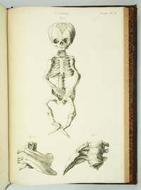
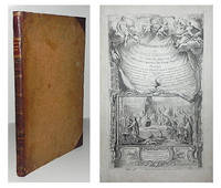
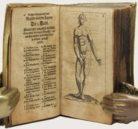
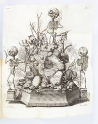
1417ST20185Amsterdam: Joannes Wolters then Jansson-Waesberg 1701-14; 1715-16. FIRST EDITIONS. 224 x 168 mm. 8 3/4 x 6 1/2". 4 p.l. 62 pp. 1 leaf errata; 2 p.l. 98 pp.; 2 p.l. 70 pp.; 1 p.l. 55 pp.; 1 p.l. 54 pp. 2 p.l. 30 pp. 1 leaf 92 pp. 2 leaves 44 pp. 4 leaves 68 pp. 8 leaves 72 pp. 1 leaf errata 12 leaves 78 pp. 10 parts in one volume. <br/> Contemporary stiff vellum smooth spine with ink titling yapp edges. WITH 42 ENGRAVED PLATES as called for by Norman six of these folding. Text in Latin and Dutch. Blake 395; Garrison-Morton 389; Heirs of Hippocrates 627; Jeremy Norman's HistoryofMedicine.com 623; Norman 1875. Vellum lightly soiled front pastedown slightly defective but the original unsophisticated binding in perfect order. Final plate with small hole due to adhesion to facing page minor damage done to image a few other trivial imperfections a couple of negligible marginal tears a faint ink stain across the top inch of text a tiny burn hole but AN UNUSUALLY BRIGHT CLEAN COPY INTERNALLY with all the important plates in exceptionally fine condition.<br/> <br/> Scarce when seen bound from its original 10 parts as here this work describes the memorable "Anatomical Treasure" of Dutch physician and anatomist Frederick Ruysch being illustrated by surrealistic charmingly macabre engravings depicting "fantastic dream-like concoctions constructed of human anatomical parts." History of Medicine Described by Norman as "probably the most original artist in the history of anatomical preparations" Ruysch 1638-1731 "enjoyed making up elaborate three-dimensional emblems of mortality from his specimens" which included fetal skeletons and preserved organs. Heirs to Hippocrates says that "the engraved illustrations deserve special mention for their whimsical almost surrealistic quality: quaintly posed skeletons surrounded by stuffed monsters strange reptiles dried plants and sea creatures." According to Norman "Ruysch's methods allowed him to prepare organs such as the liver and kidneys and keep entire corpses for years. He used a mixture of talc white wax and cinnabar for injecting vessels and an embalming fluid of alcohol made from wine or corn with black pepper added. Using his injection methods Ruysch was the first to demonstrate the occurrence of blood vessels in almost all tissues of the human body thereby destroying the Galenic belief that certain areas of the body had no vascular supply. He was also the first to show that blood vessels display diverse organ-specific patterns." Ruysch's dioramas were displayed in his home museum in Amsterdam which was open to the public; visitors included Tsar Peter the Great of Russia who purchased the collection in 1717 for the foundation of Russia's first public museum the St. Petersburg Kunstkammer. Unfortunately the Russian climate was not kind to the fragile constructions and all deteriorated over time; the dramatic plates here are the best record we have of this unique collection. Joannes Wolters, then Jansson-Waesberg unknown
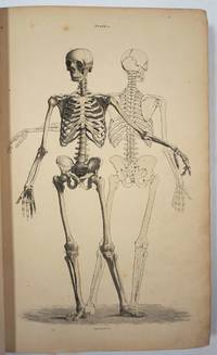
1793ST20586Lugduni Batavorum Leyden: S. & J. Luchtmans 1793 1827 1835. FIRST EDITION A COMPLETE SET. 525 x 350 mm. 20 3/4 x 14". Four volumes. <br/> First two volumes in contemporary cat's paw calf volumes III and IV in contemporary flamed calf raised bands spines gilt in compartments with central fleuron one red and one black morocco label. WITH 206 ENGRAVED ANATOMICAL PLATES all with guards. Front pastedowns with bookplate of Harvard Medical School Library; library shelf markings in white at bottom of spines; bookseller's ticket of Schönhof & Mueller Foreign Books Boston. Bibliotheca Walleriana 8549; Choulant pp. 312-13; Larousse XIX vol. 14 p. 171. Lucas L. Boer Peter L. J. Boek Andries J. van Dam and Roelof-Jan Oostra "History and highlights of the teratological collection in the Museum Anatomicum of Leiden University The Netherlands " American Journal of Medical Genetics Part A Volume 176 no. 3 2018 pp. 618-37.<br /> Shallow chips across the tail of first two volumes covers variably abraded one noticeably so corners somewhat worn partially with boards showing through vague additional signs of wear but the bindings solid and retaining much of their original appeal; plates in first two volumes with faint overall browning either because of paper stock or because of contact with guards a handful of trivial tears but all in all an extremely pleasing set the plates in volume IV very clean and bright and the plates elsewhere and the wide-margined text clean and fresh throughout.<br/> <br/> This is an extremely scarce complete set of a very substantially proportioned anatomical work with more than 200 striking giant folio engravings that emphasize birth defects and that range from clinically cheerless to heartbreaking. A physician and professor of anatomy at Leyden Eduard Sandifort 1742-1814 was commissioned by the University and the city council to publish this account of the school's anatomical collections with a focus on pathological anatomy including especially congenital abnormalities. After the publication of the first two volumes in 1793 the third and fourth were issued following Sandifort's death by his son Gerard 1779-1848 who followed in his father's professorial footsteps in Leyden. The first second and fourth volume are illustrated with copperplate engravings which Larousse tell us make this book "one of the most beautiful works of this genre" though "beautiful" needs to be understood here to mean "convincing" or "compelling" rather than aesthetically appealing. The first volume concludes with nine full-size depictions of skulls; the illustrations in the second and fourth are of a more affecting nature consisting of malformed bodies and body parts damaged or destroyed by disease and birth defects. The engravings of the conjoined twins in these volumes are especially powerful--consisting of poignant even haunting images showing undeserving victims of pronounced congenital defects though some are softened with a touch of the fanciful in their presentation. The thoroughness and accuracy of the work is so great that it remains of scholarly value even today. A recent study by Dutch curators and anatomical scholars Lucas L. Boer Peter L. J. Boek Andries J. van Dam and Roelof-Jan Oostra used "Museum Anatomicum" as a guide to identify and re-diagnose the University's historic teratological collections noting that "many historically made diagnoses could not be improved upon after re-diagnosing the specimens with contemporary dysmorphological knowledge actually confirming that these old collectors were perhaps the first dysmorphologists and can be seen as true pioneers in the field of teratology." Although single volumes and incomplete sets of this work come on the market with some regularity we have been able to trace only five complete sets at auction since 1872. And ours especially fresh and wide-margined in pleasing contemporary calf is a particularly attractive specimen. S. & J. Luchtmans unknown
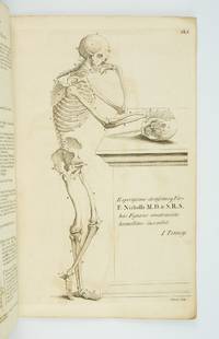
1739ST12883Leyden: Johann Arnold Langerak 1739. First Edition in Latin. 540 x 380 mm. 21 1/4 x 15". 70 leaves of text.Translated by William Dundass. <br/> Original red quarter vellum over marbled boards raised bands UNTRIMMED EDGES. Engraved printer's device on title large decorative initials and tailpieces EXTRA ENGRAVED TITLE AND 114 STRIKING PLATES OF ANATOMICAL FIGURES three folding. Wellcome II 401; Heirs of Hippocrates 468; Choulant-Frank pp. 252-3; Russell 213. ◆Vellum on spine rather worn with three one-and-one-half-inch pieces broken away revealing structure underneath paper boards quite chafed other minor problems externally but an entirely solid unrestored binding. A couple of plates with short closed marginal tears one folding plate with one-inch closed tear into image no loss untrimmed edges a little browned and brittle with isolated small chips occasional minor foxing or insignificant stains three plates lightly browned but still AN UNUSUALLY ATTRACTIVE COPY INTERNALLY with the broadest of margins with especially clean and fresh leaves and with rich impressions of the plates.<br/> <br/> With plates of notable visual impressiveness this was the finest anatomy book in England during the first half of the 18th century; it also was largely a plagiarism borrowing most of its engraved content from a work by the Dutch physician Govert Bidloo published in 1685. Bidloo's work was the first large-scale anatomical atlas to appear after Vesalius' epoch-making "De Humani Corporis Fabrica" and the plates which are highly praised by Norman and Garrison-Morton are characterized by startlingly detailed life-size depictions of the human body both adult and infant with figures flayed to reveal muscles opened to show organs and unfleshed to exhibit bones. According to Choulant-Frank Bidloo's publishers sold 300 impressions of these plates to Cowper probably to recoup some of their money after disappointing sales. Cowper took Bidloo's original 105 plates added nine of his own and produced an English translation of the original Latin text to accompany them. Discussing the original plates produced by Gerard de Lairesse 1641-1711 Norman says that the figures are displayed "in an emotional almost tender manner contrasting the raw dissected parts with the full soft surfaces of uncut flesh placing flayed bound figures in ordinary nightclothes or bedding setting ordinary household objects such as books jars or cabinets in the same scene as cut-up torsos or limbs. His illustrations brought the qualities of Dutch still-life painting into anatomical illustration and gave a new darker spiritual expression to the significance of the act of dissection." When Cowper's version of the atlas first appeared as "The Anatomy of Humane Bodies" in London in 1698 there was also a 1737 Leyden printing in English before our more scholarly Latin edition Bidloo complained to the Royal Society and accused Cowper of plagiarism and fraud resulting in much acrimony and heated pamphleteering between the two physicians. Notwithstanding this scandal Cowper's achievements and discoveries--including the pair of glands that bear his name--were considerable and his text improved significantly upon the original work. Unfortunately as the DNB notes "the notoriety of this case has served to obscure a true appreciation of Cowper and of his many original contributions to anatomical illustration." The atlases of Bidloo and Cowper appear on the market regularly but at 540 x 380 mm. the present copy is distinguished by its size which is significantly larger than what is typically seen with this edition--we have not been able to trace a copy larger than ours from marketplace or institutional records. Johann Arnold Langerak unknown
1890179459Peoria Illinois: American Manikin Company 1890. The bare bones of anatomy An educational model depicting male anatomy. It was positioned as a cheaper and more user-friendly alternative to Louis Auzoux's papier-mâché manikins themselves an accessible innovation from earlier wax models. The American Manikin represents a crucial step in the widening accessibility and popularization of anatomy and is a precursor to the mass-produced teaching models of the 20th century. The model is double-sided one face depicting muscles nerves tendons and the spinal column and the other showing muscles veins and bones. The latter face has alphabetically labelled hooks onto which illustrated steel attachments can be affixed. When all the plates are in place the model can be "dissected" by removing each layer in sequence eventually revealing the skeleton. Several of the attachments have additional diagrams on the reverse; for example the attachment illustrating the nerves of the brain also features a diagram of the inner ear on the other side. First produced in 1888 the manikin was designed by Elias Smith 1828-1913 who also published the Atlas of Colored Plates Illustrating Features of Anatomy 1889. Smith was an enterprising businessman who held patents for bicycle saddles and dental equipment. The accompanying advertisement booklet makes extensive reference to the American Manikin's superiority over Auzoux's French model. The attachment for the model's hand is illustrated with the company's calling card. The manikin was issued with 40 attachments of which 24 are present. An accompanying Teachers' Handbook was also produced not present. Wooden model approximately 1070 x 407 mm chromolithographic illustrations affixed to both faces one face with 27 metal hooks. With 24 steel attachments with similar illustrations metal stand comprising base and three attachable legs. Housed in original wooden case 1020 x 510 mm chromolithographic anatomical diagrams printed on inner panels instructional leaflet affixed to same. With 62-page advertisement booklet. Case with near-contemporary postage label of Lewis Bohnett 1880-1970 antique-collector and proprietor of "Trader Lew's Relic Museum" California; "fragile" label dated 1939 to same. Lightly worn from use lacking single hook cockling and a few chips to chromolithographic overlays colours bright; superficial wear to case: in very good condition standing firm. unknown
185729295Edinburgh: W.H. Lizars 3 St. James Square; S. Highley 32 Fleet St. London and W. Curry Junr & Co 9 Upper Sackville St. Dublin 1857. Hardcover book. Good overall. Large remarkable plates illustrating the human body skeleton organs circulatory muscular and nervous and lymphatic systems fetal respiratory eyes internal organs. The plates with tissue guards most hand colored engraved by W.H. Lizars. <br /> <br /> Owners signature on ffep "George Edward Rundle 30th September 1889" plus his bookplate on inside front cover. Rundle c. 1846 - 1906 was born in Hampshire England and received his education at the University College London and Edinburgh University qualifying in 1873. He first practiced at Hillston NSW and subsequently at Tenterfield and in Sydney by 1876. In July 1875 he was listed as enrolled on the list of medical practitioners of Tasmania in the Launceston Examiner 29 Jul 1875. He is listed in a government gazette with many degrees including a Fellow of the Royal College of Surgeons in Edinburgh in 1878. After retiring he became a trustee of the Sydney Museum and a member of the Zoological Society and for some time had been president. He died 16 Oct 1906 at 14 Wylde Street Potts Point. He is buried at Waverley Church of England cemetery. <br /> <br /> OCLC 338701549 cites 1 copy at the Univ. of York. <br /> <br /> Folio 26pp Contents 101 plates 241pp text xxxvi indices complete. Brown leather and cloth covers detached but present spine partially perished. Internally very clean. W.H. Lizars, 3 St. James Square; S. Highley 32 Fleet St. London, and W. Curry Junr & Co, 9 Upper Sackville St. Dublin hardcover
1830D7399Italy c. 1830. Hardcover. Very Good. Modern half morocco and marbled boards gilt-stamped lettering and ornament on spine; 430 x 560 mm; contains 26 anatomical drawings in red and grey chalk most of them full-page but also including double-page spreads tipped onto stubs. Spine tips and corners gently bumped; some faint dampstaining; a few leaves with repaired tears. Images of skeletal and muscular representations of arms legs feet torsos and heads. <br/><br/> hardcover
1784000285Etienne Charpentier 1784. Hardcover. See Description. Tall folio 385 mm. The entire book including the text was printed from engraved copper plates 60 plates an illustrated title page plate 29 anatomical plates and 30 calligraphic letterpress plates. Bound in a contemporary or near contemporary binding with calf spine and decorative paste paper boards - rubbed edges shelf worn old ink stain on rear cover. Both the upper portion of the spine and the tail cap are carefully restored. Board corners are neatly repaired. Interior contains an occasional minor spots of marginal foxing; a couple of tiny holes or paper defects are present on the text plate opposite plate 20. The leaves are otherwise generally clean. Francois-Michel Disdier 1708-1781 was a professor of surgery and a drawing master at the Academy of Painting in Paris. His "Tableaux anatomiques" a deluxe anatomy book for physicians was first published in 1758. Many of the plates were done by Crepy and Charpentier some after the style of Vesalius and others following Eustachi. The engraved title page designed by François Bouchet depicts students in an anatomy class studying a cadaver. The whole scene is framed in ornate baroque style. Blake NLM p. 122; Heirs of Hippocrates 894 listing 1778 edition; Hirsch II p. 190. <br/> <br/> Etienne Charpentier hardcover
174325189Leiden: Joannem & Hermannum Verbeek 1743. From the first edition of one of the greatest of all Anatomical Atlases. Tabula IX which features Albinus and Wandelaar's famous "Muscle Man" a skinned figure whose musculature is visible and defined as viewed in walking profile with the left arm raised and the right foot forward his head is turned slightly away from the viewer. Elephant folio 620 by 475 mm single folio sheet now mounted with the use of none-evasive corner tabs and protected by mylar. Very well preserved fully intact with only the most minor evidence of age. ONE OF THE MAGNIFICENT PLATES FROM "AMONG THE MOST ARTISTICALLY PERFECT OF ANATOMICAL ATLASES." Wandelaar placed his skeletons and musclemen against lush ornamental backgrounds to give them the illusion of vitality using contrasts of mass and light to produce a three-dimensional effect. The most famous plate in the atlas depicts a skeletal figure standing in front of an enormous grazing rhinoceros sketched by Wandelaar from the first living specimen in Europe which had arrived at Amsterdam zoo in 1741" Norman.<br> The plates in this large folio work and in the four supplementary works in large folio with which it is bound are unsurpassed for their cool elegant aesthetic and scientific accuracy. They were drawn and engraved by Jan Wandelaer a pupil of the engravers Jacob Fokema and Guillem van der Gouwen and the painter Gerard de Lairesse who prepared the drawings for Bidloo's atlas. Prior to working for Albinus Wandelaer worked for Friedrik Ruysch. Albinus however provided Wandelaar with the opportunity for the full expression of his talents as a draftsman and engraver. <br> In an attempt to increase the scientific accuracy of anatomical illustration Albinus and Wandelaar devised a new technique of placing nets with square webbing at specified intervals between the artist and the anatomical specimen and copying the images using the grid patterns. Wandelaer placed each figure in a carefully chosen landscape setting and the artistic results are so pleasantly successful that the anatomical figures although composed of many separate parts appear to be actually stepping out of the picture. Joannem & Hermannum Verbeek unknown
174025188Leiden: Joannem & Hermannum Verbeek 1740. From the first edition of one of the greatest of all Anatomical Atlases. Tabula V which features Albinus and Wandelaar's famous "Muscle Man" a skinned figure whose musculature is visible and defined as viewed from behind standing with the left arm raised right arm turned and his weight shifted to the right foot. Elephant folio 620 by 475 mm single folio sheet now mounted with the use of none-evasive corner tabs and protected by mylar. Very well preserved fully intact with only the most minor evidence of age. ONE OF THE MAGNIFICENT PLATES FROM "AMONG THE MOST ARTISTICALLY PERFECT OF ANATOMICAL ATLASES." Wandelaar placed his skeletons and musclemen against lush ornamental backgrounds to give them the illusion of vitality using contrasts of mass and light to produce a three-dimensional effect. The most famous plate in the atlas depicts a skeletal figure standing in front of an enormous grazing rhinoceros sketched by Wandelaar from the first living specimen in Europe which had arrived at Amsterdam zoo in 1741" Norman.<br> The plates in this large folio work and in the four supplementary works in large folio with which it is bound are unsurpassed for their cool elegant aesthetic and scientific accuracy. They were drawn and engraved by Jan Wandelaer a pupil of the engravers Jacob Fokema and Guillem van der Gouwen and the painter Gerard de Lairesse who prepared the drawings for Bidloo's atlas. Prior to working for Albinus Wandelaer worked for Friedrik Ruysch. Albinus however provided Wandelaar with the opportunity for the full expression of his talents as a draftsman and engraver. <br> In an attempt to increase the scientific accuracy of anatomical illustration Albinus and Wandelaar devised a new technique of placing nets with square webbing at specified intervals between the artist and the anatomical specimen and copying the images using the grid patterns. Wandelaer placed each figure in a carefully chosen landscape setting and the artistic results are so pleasantly successful that the anatomical figures although composed of many separate parts appear to be actually stepping out of the picture. Joannem & Hermannum Verbeek unknown
174025187Leiden: Joannem & Hermannum Verbeek 1740. From the first edition of one of the greatest of all Anatomical Atlases. Tabula V which features a human skeleton in a three-quarter view and seen from the rear is portrayed in a walking motion with the left hand raised and extended. Elephant folio ca. 620 by 475 mm single folio sheet now mounted with the use of non-evasive corner tabs and protected by mylar. Very well preserved fully intact with only the most minor evidence of age. ONE OF THE MAGNIFICENT PLATES FROM "AMONG THE MOST ARTISTICALLY PERFECT OF ANATOMICAL ATLASES." Wandelaar placed his skeletons and musclemen against lush ornamental backgrounds to give them the illusion of vitality using contrasts of mass and light to produce a three-dimensional effect. The most famous plate in the atlas depicts a skeletal figure standing in front of an enormous grazing rhinoceros sketched by Wandelaar from the first living specimen in Europe which had arrived at Amsterdam Zoo in 1741" Norman.<br> The plates in this large folio work and in the four supplementary works in large folio with which it is bound are unsurpassed for their cool elegant aesthetic and scientific accuracy. They were drawn and engraved by Jan Wandelaer a pupil of the engravers Jacob Fokema and Guillem van der Gouwen and the painter Gerard de Lairesse who prepared the drawings for Bidloo's atlas. Prior to working for Albinus Wandelaer worked for Friedrik Ruysch. Albinus however provided Wandelaar with the opportunity for the full expression of his talents as a draftsman and engraver. <br> In an attempt to increase the scientific accuracy of anatomical illustration Albinus and Wandelaar devised a new technique of placing nets with square webbing at specified intervals between the artist and the anatomical specimen and copying the images using the grid patterns. Wandelaer placed each figure in a carefully chosen landscape setting and the artistic results are so pleasantly successful that the anatomical figures although composed of many separate parts appear to be actually stepping out of the picture. Joannem & Hermannum Verbeek unknown
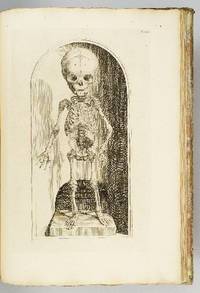
1770ST20219London: Printed for Robert Sayer ca. 1770. 380 x 242 mm. 15 x 9 1/2". 4 leaves of text followed by plates. <br/> Original gray paper wrappers. WITH EIGHT FINE ENGRAVED PLATES of the male body in various poses three depicting the skeleton and five the musculature. Russell 816; Wellcome V p. 273; ESTC N51161. See also Russell "John Tinney's Compendium Anatomicum and its publishers." Wrapper a bit soiled and foxed corners torn with two snags two-inch and half-inch to lower cover most of spine chipped away but the stitching holding the book firmly together; mild offsetting from engravings and a few spots of foxing but a really excellent copy internally the leaves fresh and clean and with very good impressions of the engravings.<br/> <br/> This rare collection of striking anatomical engravings is wonderfully preserved in its original unrestored wrappers. First published in 1743 as "Compendium Anatomicum" the work's plates are adapted from the famous anatomies of Vesalius and Cowper. It was intended as a reference work for artists but as its name suggests it was published with a wider audience in mind. As Tinney humbly tells the reader in the subtitle this is "a work not only very useful but absolutely necessary to painters statuaries and all professors of drawing and design as well as a proper introduction to the study of anatomy for the use of young surgeons" not to mention an "instructive furniture for the studies and libraries of the curious." The work remained in print for a full century though because it was a frequently used book few copies have come down to us in collectible condition. John Tinney ca. 1706-61 was an engraver and print seller who dealt in a wide variety of material being particularly known for maps and satirical prints as well as the present work. Our edition while undated almost certainly dates to the period between 1762-70. The first posthumous edition of "A Compendious Treatise" was released in 1762 by Robert Sayer a map and print seller who was an associate of Tinney's during his lifetime and who likely purchased his stock of plates from his widow following his 1761 death. In 1770 Sayer began a partnership with John Bennett after which the firm was known as "Sayer and Bennett." Since the present edition was published under Sayer's name alone it no doubt comes from the period following the 1762 edition but before the partnership. The present copy has made it through the centuries in remarkably good condition given its flimsy binding and the plates inside with their cheerfully macabre figures remain quite fresh and pleasing. Printed for Robert Sayer unknown
1796015628Cambridge: Printed by J. Burges Printer to the University; And Sold By W. H. Lunn and J. Deighton Cambridge; Messrs. White Fleet-Street and J. Walter Charing-Cross London; And J. Cook Oxford 1796. First Edition First Impression . Cloth Bound Boards. Very Good. 1796 First Edition. Size slim square quarto 72 pages plus 15 pages of plates at the rear. Bound in modern brown cloth Birdsall & Son of Northampton with gilt titles and vignette to the spine. Condition very good corners and spine ends a little rubbed some light spotting and dust soiling at front and rear pages including to the margins of the final plate else the contents are very clean throughout. Contains 15 full page engraved plates at the rear of the book. The plates depict skulls and other bones in the head of various different animals and birds. The title states volume I This was intended as a projected work in two volumes of five fascicules but this first fascicule was the only one ever published. Harwood was a professor of anatomy at Cambridge and an early developer of blood transfusion techniques. OCLC lists only ten copies and no copies sold at auction have been traced. Size: 4to - over 9" - 12" tall <br/> <br/> Printed by J. Burges Printer to the University; And Sold By W. H. Lunn, and J. Deighton Cambridge; Messrs. White, Fleet-Street, hardcover
17341408797Paris: Guillaume Cavelier 1734. Later Editions. Hardcover. Octavos Four Volumes bound as two. The first book contains a single volume; the second book contains the second volume of Anatomie Chirurgicale in addition to two other standalone works. In Good condition. In full brown leather bindings; spines paneled with elaborate gilt decoration and gilt titling on tan leather labels; gilt roll to board edges. Scuffing and wear to boards with peeling to covers. All edges and fore corners rubbed with sections of exposed board. Chipping to heads and tails of spines; heaviest at head of Vol. I with head band nearly detached. Text block edges show shelf wear and dust soiling. Lightly age toned and very sparsely foxed throughout. Splitting to hinges. Rear pastedown in Vol. I and both pastedowns in Vol. II are separating from the boards. ~2 in. closed tear in Vol. I p. 39. Splitting in gutters of several signatures throughout both volumes due to damage to end bands. Closed tears affect some fold-out plates. Tidemarks affect pages in the rear of both volumes.<br /> <br /> <br> <br> <br /> <br /> CONTENTS: Anatomie Chirurgicale Vol. I xxiv 514 2 pages plus plates i-xxx; Anatomie Chirurgicale Vol. II viii 313 336-352 453-465 366-403 pages plus plates xxxi-xlviii bound together with Observations Anatomiques viii 175 pages plus 8 plates and Six Observations 2 36 2 pages. Shelved in Room A. The first work in this set is the second edition of Flemish surgeon Jean Palfin's Jan Palfijn's very famous treatise on anatomy which was first published in 1726 under the title Anatomie du Corps Humain. This work and its earlier edition were very influential in the medical field for their detailed anatomical illustrations and descriptive actionable advice for surgeons becoming a widespread authoritative medical text throughout Europe and other parts of the world. <br /> <br /> <br> <br> <br /> <br /> The second work bound together with Vol. II of Anatomie Chirurgicale is a French translation of Frederik Ruysch's Observationum Anatomico-Chirurgicarum Centuria. Ruysch was a renowned anatomist and botanist in his own right most well-known for his anatomical museum - the Cabinet of Curiosities - and his improved embalming technique which preserved bodies for longer periods of time in a more visually life-like state.<br /> <br /> <br> <br> <br /> <br /> The final work also bound together with Vol. II of Anatomie Chirurgicale and Observations Anatomiques is a revised edition of Six Observations by Michel Brisseau who is notable for being the first to demonstrate the true nature of cataracts as an increased opacity of the lens of the eye rather than a film that covered the eye which was the prevailing belief at the time. Together these works comprise a collection of highly influential studies treatises and observations on anatomy and surgery by some of the most notable medical practitioners of the late 17th and early 18th centuries. 1408797. Special Collections. Guillaume Cavelier hardcover
182840962London 1828. <p>Parliament. House of Commons. Report from the select committee on anatomy. Folio. 150pp. London: House of Commons 22 July 1828. 331 x 212 mm. 19th century boards rebacked and recornered in calf light edgewear. Very good copy. Old medical library stamp on title and first page.</p> <p>First Edition of this highly interesting and entertaining report on the British body-snatching crisis. Since the mid-eighteenth century obtaining cadavers for teaching purposes had been regulated in Britain by the Murder Act of 1752 which stipulated that only the corpses of executed criminals could be used for dissection. By the beginning of the nineteenth century however improvements in medical science coupled with a substantial drop in the number of executions caused the demand for cadavers to far outstrip the legal supply. This situation was ripe for exploitation by "resurrection men" criminals who robbed the graves of the newly deceased and sold their corpses to teachers of anatomy who of necessity turned a blind eye to the illegality of these transactions. Some grave-robbers even resorted to murder including the infamous William Burke who in December 1828 was arrested in Edinburgh for the murders of over a dozen victims whose corpses he and his partner Hare sold to an anatomical demonstrator connected to Edinburgh University.</p> <p>In the first half of 1828 in response to increasing calls for reform the British Parliament appointed a committee to "enquire into the manner of obtaining subjects for dissection by schools of Anatomy and the State of law affecting persons employed in obtaining and dissecting bodies." During the course of its investigation the committee heard testimony from a wide range of witnesses from eminent medical men to procurers of bodies for medical schools these last identified only by initials. The medical men included Sir Astley Cooper Benjamin Collins Brodie John Abernethy William Lawrence Herbert Mayo Granville Sharp Pattison who himself was indicted for body-snatching at the age of 23 Thomas Southwood Smith Henry Halford John Webster and Benjamin Harrison the treasurer of Guy's Hospital. The witness list can be found on page 13 of the committee's report. The testimony of these men reproduced in full in the report is followed by several appendices including tables of paupers' deaths broken down by parish; the committee was proposing legislation that would allow the state to seize unclaimed corpses from workhouses and sell them to surgical schools. The committee's efforts were successful: In 1832 Parliament passed the Anatomy Act granting licenses to teachers of anatomy and giving physicians surgeons and medical students legal access to corpses unclaimed after death. Wise The Italian Boy: A Tale of Murder and Body Snatching in 1830s London 2004. Garrison-Morton.com 7096.</p> . unknown
170434368Leipzig: Thomas Fritsch 1704. First German Edition. Illustrated with an engraved frontispiece of the author and 31 additional full-page engravings depicting various anatomical views. 8vo bound in the original contemporary vellum the spine with calligraphic titling by hand. viii 742 26 Register pp. A very good copy the binding still strong and well preserved the text with the usual mellowing and aging still crisp and very usable. FIRST GERMAN EDITION of this important work on anatomy partnered with the work of Philippi Verheyen Anmerckungen In Die Anatomiam Blancardi. Brieff An den weitberühmten Hrn. Friedrich Ruyschium.<br> 'Verheyen was a prolific writer including several books and manuscripts. His main work published originally in Latin the “Corporis Humani Anatomiae†was translated into multiple languages as here. This book became one of the most used anatomy books of the time.<br> The following excerpt of his book "Corporis Humani Anatomiae Liber Primus" reads : " . QuintusAuricularis quia cum minimum sit auribus expurgandis est aptissimus" translates as".the fifth finger called Auricularis because how small it is is most suitable to clean the ears". Incredibly anatomists at that time called the fifth digit "digitus auricularis".<br> Verheyen is credited with the creation of the eponym the “Achilles tendon†which denominates the common tendon for the gastrocnemius and soleus muscle although at the time he called it the “Chorda Achillisâ€. He also described the kidneys in detail especially the arterial “stars†found on the surface of the kidney which are today known as the “Stars of Verheyen†'. E. A. Miranda 2014 M.T.D. Thomas Fritsch hardcover
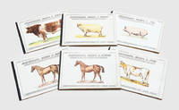
1900188515London: Vinton & Company Ltd c.1900. A set of elaborate moveable books depicting the anatomy of farmyard animals. Each includes five layers of folding colour diagrams showing the skeleton arteries and veins muscles and organs accompanied by a key. It was printed in Bavaria a prestigious centre of moveable books and home to skilled artisanal paper engineers such as Ernest Nister and Lothar Meggendorfer. This is the second edition of the set. Six works oblong quarto. Each 8 pp. and with folding chromolithographic illustration comprising two large overslips and multiple lift-the-flap tabs representing internal anatomy. Original printed boards black cloth backstrips wire-stitched as issued. Foxing toning and soiling to boards and contents wear to backstrips wire-stitches rusted but holding firm outer margins of contents with a few chips and damp stains a handful of neat repairs to folding stubs colours bright: in very good condition. hardcover
BN66051Anatomie des Auges DOZ and Maidowsky Werner <br/><br/> unknown
61852Printed by J. Dixwell No. 148 St. Martin's Lane near Charing Cross. London. 1777. 8vo. pp. 8168 i182-250. Text is continuous despite erratic pagination. Contemporary half calf and marbled boards though now with a later cloth spine occasional foxing more especially at the front ex-libris with stamps and book-label on the front paste-down neat oval stamp on verso of title and in margin of last page overall a very good copy. MAGNUS FALCONAR not in Munk or Plarr was a friend and colleague of William Hewson surgeon anatomist and physiologist; in 1772 Hewson established and ran an anatomy school at 36 Craven Street where Franklin lodged in London now the Benjamin Franklin House museum. In 1998 workmen restoring this building dug up the remains of six children and four adults hidden below the home. The Times reported on 11 February 1998: 'Initial estimates are that the bones are about 200 years old and were buried at the time Franklin was living in the house which was his home from 1757 to 1762 and from 1764 to 1775. Most of the bones show signs of having been dissected sawn or cut. One skull has been drilled with several holes. Paul Knapman the Westminster Coroner said yesterday: "I cannot totally discount the possibility of a crime. There is still a possibility that I may have to hold an inquest." ' The Friends of Benjamin Franklin House noted that the bones were likely placed there by Hewson who lived in the house for two years. They also note that Franklin likely knew what Hewson was doing. Proof was demonstrated by archaeological evidence which showed liquid mercury associated with turtle bones and vermilion colouring associated with dog bones found in the deposit. Hewson had documented experimentation on the lymphatic system using both substances and animals. Hewson died on 1 May 1774 as a result of sepsis contracted whilst dissecting a cadaver. His work was continued after his death by Magnus Falconar who had married Hewson's sister Dorothy in September 1774. Falconar repeated Hewson's experiments on the spleen and thymus and as a result re-published Hewson's work on red blood cells in 1777 together with his corroboration Experimental inquiries: part the third. Containing a description of the red particles of the blood in the human subject and in other animals; with an account of the structure and offices of the lymphatic glands of the Thymus Gland and of the Spleen: being the remaining part of the observations and experiments of the late Mr. William Hewson F. R. S. and Teacher of Anatomy. By Magnus Falconar Surgeon and Teacher of Anatomy. Printed for T. Longman No. 39. Pater-Noster-Row MDCCLXXVII. Samuel Paterson in 1778 published the catalogue of the auction sale of Falconar's anatomical collection - 'Museum falconarianum. A catalogue of the entire and capital museum of anatomical preparations and other subjects of natural history; a great variety of chirurgical anatomical and philosophical instruments; medicaments cabinets preparation-glasses and other effects; of the late Mr. Magnus Falconar surgeon and professor of anatomy deceased: which by order of the adminstrator will be sold by auction by Mr. Paterson at his Great Room No 6. in King-Street Covent-Garden London on Monday the 12th of October 1778 and the nine following evenings to begin precisely at five o'clock. To be viewed on Wednesday the 7th instant and to the time of sale. Catalogues price one shilling may be had at the place of sale; where also may be had Mr. Falconar's synopsis of his course of lectures on anatomy and surgery printed only for the use of his pupils and never before published Price five shillings.' ESTC also records the publication of 'A syllabus of a course of lectures on anatomy on physiology and on the operations and practice of surgery' 1777 Printed by J. Dixwell, No. 148, St. Martin's Lane, near Charing Cross. London. 1777. 8vo. hardcover
BN66125Allgemeine Anatomie und Bewegungssystem 1 DVD-ROMFür Windows 98 SE/2000/ME/XP/Vista <br/><br/> unknown
BN90792Thieme. Klinische Anatomie der Halswirbelsäule Hardcover <br/><br/>Klinische Anatomie der Halswirbelsäule Hardcover Klinische Anatomie der Halswirbelsäule Hardcover Thieme hardcover
SONG1878576488McGraw-Hill Education 0000-00-00. cards. Used: Good. 4.75x6.25x6.00. Buy with confidence. Excellent Customer Service & Return policy. McGraw-Hill Education unknown
BN91163Schober. Farbatlas der Anatomie der Ratte. Sektionsanleitung Hardcover <br/><br/>Farbatlas der Anatomie der Ratte. Sektionsanleitung Hardcover Farbatlas der Anatomie der Ratte. Sektionsanleitung Hardcover Schober hardcover
BN91390Parey bei Blackwell. Lehrbuch der Anatomie der Haustiere 5 Bde. Bd.2 Eingeweide Frehwein Josef; Gasse Hagen; Leiser Rudolf; Nickel Richard; Schummer August and Seiferle Eugen <br/><br/>Lehrbuch der Anatomie der Haustiere 5 Bde. Bd.2 Eingeweide Frehwein Josef; Gasse Hagen; Leiser Rudolf; Nickel Richard; Schummer August and Seiferle Eugen Lehrbuch der Anatomie der Haustiere 5 Bde. Bd.2 Eingeweide Frehwein Josef; Gasse Hagen; Leiser Rudolf; Nickel Richard; Schummer August and Seiferle Eugen Parey bei Blackwell unknown
BN66422Organe des aktiven Bewegungsapparates der Koordination der Umweltbeziehung des Stoffwechsels Vergleichende Anatomie der Wirbeltiere Bd.3: Band . des Stoffwechsels und der Fortpflanzung Starck D. <br/><br/> unknown
BN66626Anatomie des Kiefer-Gesichts-Bereiches Schumacher Gert-Horst <br/><br/> unknown