5 849 résultats
482Rome, Antonii de Rubeis, 1741, un volume in folio, (42,20 cm x 30 cm) relié en plein parchemin, dos muet (habiles restaurations sur les plats, quelques traces de poussière dans les marges de la dernière page de texte), 2 feuillets non chiffrés (titre+avis au lecteur), 84pp., 27 SUPERBES PLANCHES
1403Lugduni batavorum, Joannem Arnoldum Langerak, 1739, un fort volume grand in folio (535x365mm,leaf 525x355mm) relié en PLEIN MAROQUIN ROUGE, (reliure de l'époque), dos orné de fers dorés, toutes tranches dorées, filets dorés sur les plats, 9 ff. (titre gravé, titre imprimé en noir en en rouge, dedicace...), 59 ff. (explication des planches), 2 ff (index), 114 PLANCHES (105 pl. numérotées 1 à 105 + 9 planches numérotées 1 à 9
1417ST20185Amsterdam: Joannes Wolters then Jansson-Waesberg 1701-14; 1715-16. FIRST EDITIONS. 224 x 168 mm. 8 3/4 x 6 1/2". 4 p.l. 62 pp. 1 leaf errata; 2 p.l. 98 pp.; 2 p.l. 70 pp.; 1 p.l. 55 pp.; 1 p.l. 54 pp. 2 p.l. 30 pp. 1 leaf 92 pp. 2 leaves 44 pp. 4 leaves 68 pp. 8 leaves 72 pp. 1 leaf errata 12 leaves 78 pp. 10 parts in one volume. <br/> Contemporary stiff vellum smooth spine with ink titling yapp edges. WITH 42 ENGRAVED PLATES as called for by Norman six of these folding. Text in Latin and Dutch. Blake 395; Garrison-Morton 389; Heirs of Hippocrates 627; Jeremy Norman's HistoryofMedicine.com 623; Norman 1875. Vellum lightly soiled front pastedown slightly defective but the original unsophisticated binding in perfect order. Final plate with small hole due to adhesion to facing page minor damage done to image a few other trivial imperfections a couple of negligible marginal tears a faint ink stain across the top inch of text a tiny burn hole but AN UNUSUALLY BRIGHT CLEAN COPY INTERNALLY with all the important plates in exceptionally fine condition.<br/> <br/> Scarce when seen bound from its original 10 parts as here this work describes the memorable "Anatomical Treasure" of Dutch physician and anatomist Frederick Ruysch being illustrated by surrealistic charmingly macabre engravings depicting "fantastic dream-like concoctions constructed of human anatomical parts." History of Medicine Described by Norman as "probably the most original artist in the history of anatomical preparations" Ruysch 1638-1731 "enjoyed making up elaborate three-dimensional emblems of mortality from his specimens" which included fetal skeletons and preserved organs. Heirs to Hippocrates says that "the engraved illustrations deserve special mention for their whimsical almost surrealistic quality: quaintly posed skeletons surrounded by stuffed monsters strange reptiles dried plants and sea creatures." According to Norman "Ruysch's methods allowed him to prepare organs such as the liver and kidneys and keep entire corpses for years. He used a mixture of talc white wax and cinnabar for injecting vessels and an embalming fluid of alcohol made from wine or corn with black pepper added. Using his injection methods Ruysch was the first to demonstrate the occurrence of blood vessels in almost all tissues of the human body thereby destroying the Galenic belief that certain areas of the body had no vascular supply. He was also the first to show that blood vessels display diverse organ-specific patterns." Ruysch's dioramas were displayed in his home museum in Amsterdam which was open to the public; visitors included Tsar Peter the Great of Russia who purchased the collection in 1717 for the foundation of Russia's first public museum the St. Petersburg Kunstkammer. Unfortunately the Russian climate was not kind to the fragile constructions and all deteriorated over time; the dramatic plates here are the best record we have of this unique collection. Joannes Wolters, then Jansson-Waesberg unknown
182343552Paris: C. de Lasteyrie 1823. <p>The Double Elephant Folio of Anatomy </p> <p>Mascagni Paolo 1752-1815. Antommarchi Francesco 1789-1838 ed. Planches anatomiques du corps humain executes d'après les dimensions naturelles . . . Double elephant folio atlas. Lithographed part-title leaf title leaf and 83 lithographed plates 48 black and white 35 outline by Charles-Philibert de Lasteyrie 1759-1849 all leaves folded and mounted on guards. Paris: C. de Lasteyrie 1823- 1826. 623 x 509 mm. individual leaves measure 907 x 623 mm. / 35.6875 x 24.5 inches unfolded. 20th century half morocco paste paper boards light wear. Discreet repairs to margins of part-title minor occasional foxing but very good.</p> <p> Considering that it is among the rarest of all anatomies and certainly the largest it is remarkable that two nearly identical editions of Mascagni's posthumous life-size anatomy were published almost simultaneously. The present lithographed edition was issued between 1823 and 1826. An edition with engraved plates was also published in Pisa under the title Anatomia universa 1823-32. Though the two editions were printed by different processes the image quality of the two is remarkably similar and it is debatable which is superior from either the artistic or scientific standpoint. Antommarchi's version in an homage to Vesalius includes imaginary landscape backgrounds created for the base of his musclemen; these do not appear in the Italian edition. There are other subtle differences. Antommarchi included letter keys within the images of some of the less-complex plates eliminating the need for outline plates to those images. He also published more anatomical plates than the Italian edition.</p> <p> The publication history of these two editions is complex and usually misunderstood. The edition we are offering was issued in 15 parts between 1823 and 1826 by the lithographic press of the Comte de Lasteyrie one of the two founders of lithography in France an accompanying text volume not present with this copy was issued in 1826 and bears the imprint of Lasteyrie's successor R. Brégeaut. The atlas with magnificent plates printed on single broadsheets measuring 970 x 650 mm. uncut is comparable in size to the double elephant folio edition of Audubon's Birds of America 1827-38 which measures about 985 x 660 mm. It is without doubt the largest lithographically printed book issued during the incunabula period of lithography. The atlas was issued in both uncolored and colored versions; according to the part-title included with this copy uncolored fascicles sold for 30 francs each and colored ones for 80 francs each. Choulant writing in the 1840s when copies of both editions might have remained available from the publishers states that copies of the completed version with colored plates could be purchased for 150 francs and uncolored copies for 375 francs. Because the plates are so large in some extant copies of the atlas they are backed with linen and cut for folding with some resulting loss of image. This is not the case here: Each plate is folded horizontally and mounted on a guard preserving the entire image.</p> . C. de Lasteyrie unknown books
1793ST20586Lugduni Batavorum Leyden: S. & J. Luchtmans 1793 1827 1835. FIRST EDITION A COMPLETE SET. 525 x 350 mm. 20 3/4 x 14". Four volumes. <br/> First two volumes in contemporary cat's paw calf volumes III and IV in contemporary flamed calf raised bands spines gilt in compartments with central fleuron one red and one black morocco label. WITH 206 ENGRAVED ANATOMICAL PLATES all with guards. Front pastedowns with bookplate of Harvard Medical School Library; library shelf markings in white at bottom of spines; bookseller's ticket of Schönhof & Mueller Foreign Books Boston. Bibliotheca Walleriana 8549; Choulant pp. 312-13; Larousse XIX vol. 14 p. 171. Lucas L. Boer Peter L. J. Boek Andries J. van Dam and Roelof-Jan Oostra "History and highlights of the teratological collection in the Museum Anatomicum of Leiden University The Netherlands " American Journal of Medical Genetics Part A Volume 176 no. 3 2018 pp. 618-37.<br /> Shallow chips across the tail of first two volumes covers variably abraded one noticeably so corners somewhat worn partially with boards showing through vague additional signs of wear but the bindings solid and retaining much of their original appeal; plates in first two volumes with faint overall browning either because of paper stock or because of contact with guards a handful of trivial tears but all in all an extremely pleasing set the plates in volume IV very clean and bright and the plates elsewhere and the wide-margined text clean and fresh throughout.<br/> <br/> This is an extremely scarce complete set of a very substantially proportioned anatomical work with more than 200 striking giant folio engravings that emphasize birth defects and that range from clinically cheerless to heartbreaking. A physician and professor of anatomy at Leyden Eduard Sandifort 1742-1814 was commissioned by the University and the city council to publish this account of the school's anatomical collections with a focus on pathological anatomy including especially congenital abnormalities. After the publication of the first two volumes in 1793 the third and fourth were issued following Sandifort's death by his son Gerard 1779-1848 who followed in his father's professorial footsteps in Leyden. The first second and fourth volume are illustrated with copperplate engravings which Larousse tell us make this book "one of the most beautiful works of this genre" though "beautiful" needs to be understood here to mean "convincing" or "compelling" rather than aesthetically appealing. The first volume concludes with nine full-size depictions of skulls; the illustrations in the second and fourth are of a more affecting nature consisting of malformed bodies and body parts damaged or destroyed by disease and birth defects. The engravings of the conjoined twins in these volumes are especially powerful--consisting of poignant even haunting images showing undeserving victims of pronounced congenital defects though some are softened with a touch of the fanciful in their presentation. The thoroughness and accuracy of the work is so great that it remains of scholarly value even today. A recent study by Dutch curators and anatomical scholars Lucas L. Boer Peter L. J. Boek Andries J. van Dam and Roelof-Jan Oostra used "Museum Anatomicum" as a guide to identify and re-diagnose the University's historic teratological collections noting that "many historically made diagnoses could not be improved upon after re-diagnosing the specimens with contemporary dysmorphological knowledge actually confirming that these old collectors were perhaps the first dysmorphologists and can be seen as true pioneers in the field of teratology." Although single volumes and incomplete sets of this work come on the market with some regularity we have been able to trace only five complete sets at auction since 1872. And ours especially fresh and wide-margined in pleasing contemporary calf is a particularly attractive specimen. S. & J. Luchtmans unknown
4394P., BAILLIERE, 1837-1841, 3 VOLUMES in 8 de TEXTE et 1 ATLAS GRAND IN FOLIO D'ATLAS, reliés en demi-basane rouge dos ornés de fers de filets dorés,T.1 (texte): 16pp. (compris faux-titre+titre), 638pp., 6 PLANCHES hors texte, T.2 (texte) : (2-faux titre + titre), 620pp., T.3 (texte) : (2-faux-titre + titre) 810pp. - ATLAS : 3 feuillets non chiffrés (faux titre+titre+dédicace), 4pp. (table), 43 feuillets non chiffrés d'explication des planches (soit 86pp. au total), 60 MAGNIFIQUES PLANCHES EN COULEURS
in-folio (mm.300x200, ff.12 nn., pp.442, ff.14 nn., titolo inciso con figure allegoriche e ritratto dell'autore, al recto del f.di dedica ritratto di Enrico IV con le figure della Giustizia e della Prudenza; 26 stupende figure anatomiche, assai dettagliate, inc . a piena pagina, gran numero di capilettera istoriati in silografia su otto linee d'altezza. Testo in elegante carattere tondo e corsivo. Legatura coeva in pieno cuoio bruno, dorso a nervi con titolo e ricchi fregi oro, in perfetto stato. Seconda edizione (prima Paris 1599). E' uno dei più importanti trattati anatomici e medici del tardo cinquecento e le belle figure si ispirano ai rami di Vesalio, Valverde, Coiter ed altri. Il Du Laurens (1558-1609) fu medico di corte di Enrico IV e Maria de Medici e successivamente capo della prestigiosa facoltà di medicina a Montpellier. Per primo, tra l'altro, descrisse con precisione l'arteria polmonare e la circolazione del sangue in generale. Esemplare assai bello, puro e fresco.. Choulant p.222. Cushing n.117. Durling 1312. Wellcome 1917. Bird 774..
17255551Leiden, J. du Vivie et J. & H. Verbeek, 1725. 2 Bde. 21 Bll., 572 S.; 4 Bll.; S. 577 - 684, 1 Bl., S. 685 - 1156, 26 Bll. mit 2 wdh. Kupfer-Titelvignetten und 38 Textkupfern. 1 Kupfertitel und 82 (12 gefalt.) Kupfertafeln (es fehlt das Kupfer-Porträt). Gr.-Fol. Mod. HLdr. auf 5 Bünden mit Rückentitel und modernen Buntpapierbezügen ((etw. berieben). [6 Warenabbildungen]
Questa straordinaria opera di anatomia è composta da: - un vol. di testo in folio (mm. 510x330), mz. pelle mod. con ang., dorso a cordoni con tit. oro: “Explication des planches anatomiques du corps humain.. par le Docteur F. Antonmarchi”, pp. (6),VII, V-VIII,288.<br> - una raccolta di tavole in folio max. (mm. 635x410) contenuta in un solido astuccio (spacchi ai bordi) in mz. pelle mod. con legacci, dorso a cordoni con tit. oro. Come da dettagliata descriz. nel vol. di testo, le tavole - tutte a doppia pag. - sono così ripartite: 48 sono in chiaroscuro e 35 sono ripetute al tratto, ciasc. con una o più figure, disegnate da Pedretti e pregevolmente litografate; l’importante collezione è introdotta da un frontespizio inquadrato in cornice floreale (sempre a doppia pag.). "Edizione originale", molto rara trovarla completa.<br> “Antonmarchi served as personal physician to Napoleon at St. Helena and achieved a certain editorial notoriety as the editor of Paolo Mascagni’s posthumous "Prodromo della grande anatomia". Upon termination of his obligations with Mascagni’s heirs, he seems to have considered the rights his own and republished the plates in the present edition, with some modifications, under his own name - without denying their provenance. He nevertheless failed to cite Mascagni on the title page and this was poorly received by the medical community. The work was conceived so that the entire human body might be represented (in life size) by joining three of the plates together. This may be achieved a number of times to depict the complete skeleton, the blood vessels, the muscles (front and back) and the viscera. Further detailed plates are dedicated to the brain, the heart, the lymphatic system, etc.”. Cfr. Choulant “History and bibliography of anatomic illustration”, pp. 319-320 che cita erroneam. 45 + 45 tavv. - Hirsch,I, p. 154-55 con 83 tavv. come il ns. esemplare. <br> Il vol. di testo: occhietto restaur. per strappi, alone al marg. super., fiorit. o arross. interc. nel t. - Le tavv.: prime 12 con alone al marg. inf., altre 20 ca. più o meno fiorite, ma certam. un buon esemplare.
1739ST12883Leyden: Johann Arnold Langerak 1739. First Edition in Latin. 540 x 380 mm. 21 1/4 x 15". 70 leaves of text.Translated by William Dundass. <br/> Original red quarter vellum over marbled boards raised bands UNTRIMMED EDGES. Engraved printer's device on title large decorative initials and tailpieces EXTRA ENGRAVED TITLE AND 114 STRIKING PLATES OF ANATOMICAL FIGURES three folding. Wellcome II 401; Heirs of Hippocrates 468; Choulant-Frank pp. 252-3; Russell 213. ◆Vellum on spine rather worn with three one-and-one-half-inch pieces broken away revealing structure underneath paper boards quite chafed other minor problems externally but an entirely solid unrestored binding. A couple of plates with short closed marginal tears one folding plate with one-inch closed tear into image no loss untrimmed edges a little browned and brittle with isolated small chips occasional minor foxing or insignificant stains three plates lightly browned but still AN UNUSUALLY ATTRACTIVE COPY INTERNALLY with the broadest of margins with especially clean and fresh leaves and with rich impressions of the plates.<br/> <br/> With plates of notable visual impressiveness this was the finest anatomy book in England during the first half of the 18th century; it also was largely a plagiarism borrowing most of its engraved content from a work by the Dutch physician Govert Bidloo published in 1685. Bidloo's work was the first large-scale anatomical atlas to appear after Vesalius' epoch-making "De Humani Corporis Fabrica" and the plates which are highly praised by Norman and Garrison-Morton are characterized by startlingly detailed life-size depictions of the human body both adult and infant with figures flayed to reveal muscles opened to show organs and unfleshed to exhibit bones. According to Choulant-Frank Bidloo's publishers sold 300 impressions of these plates to Cowper probably to recoup some of their money after disappointing sales. Cowper took Bidloo's original 105 plates added nine of his own and produced an English translation of the original Latin text to accompany them. Discussing the original plates produced by Gerard de Lairesse 1641-1711 Norman says that the figures are displayed "in an emotional almost tender manner contrasting the raw dissected parts with the full soft surfaces of uncut flesh placing flayed bound figures in ordinary nightclothes or bedding setting ordinary household objects such as books jars or cabinets in the same scene as cut-up torsos or limbs. His illustrations brought the qualities of Dutch still-life painting into anatomical illustration and gave a new darker spiritual expression to the significance of the act of dissection." When Cowper's version of the atlas first appeared as "The Anatomy of Humane Bodies" in London in 1698 there was also a 1737 Leyden printing in English before our more scholarly Latin edition Bidloo complained to the Royal Society and accused Cowper of plagiarism and fraud resulting in much acrimony and heated pamphleteering between the two physicians. Notwithstanding this scandal Cowper's achievements and discoveries--including the pair of glands that bear his name--were considerable and his text improved significantly upon the original work. Unfortunately as the DNB notes "the notoriety of this case has served to obscure a true appreciation of Cowper and of his many original contributions to anatomical illustration." The atlases of Bidloo and Cowper appear on the market regularly but at 540 x 380 mm. the present copy is distinguished by its size which is significantly larger than what is typically seen with this edition--we have not been able to trace a copy larger than ours from marketplace or institutional records. Johann Arnold Langerak unknown
in-folio (315x205 mm.), ff.(18), 154. Legatura antica piena pergamena, dorso rifatto, con titolo e fregi oro. Frontispizio inciso entro bella bordura a motivo architettonico con figure anatomiche, grandi iniziali istoriate. Classica opera, illustrata da 42 belle figure anatomiche a piena pagina nel testo, disegnate da Gaspar Becerra e incise in rame da Nicolas Beatrizet, le medesime già apparse nell'edizione spagnuola del 1556. Il testo è diviso in sette libri o sezioni (ossa, muscoli, vene, nervi, etc.) ed è ricco di indici e richiami. Prima edizione italiana, rara prima tiratura con data 1558, tradotta da Anton Tabo con la supervisione di Valverde, assai stimata e importante in campo scientifico-medico, illustrata con belle tavole ispirate da quelle del Vesalio e migliorate rispetto a quelle. Valverde, la cui opera apparve in spagnolo nel 1556, è considerato uno dei più importanti anatomisti del XVI secolo. Esemplare assai bello, con ampi margini.. Cushing VI-D-35. Mortimer n.513:( Tiratura del 1560). Wellcome 6476 (id.). Choulant-Frank 205-208..
19078602W. Engelmann, Leipzig 1849-1907. durchschnittlich ca. 650 S. je Jahrgang mit sehr zahlreichen zum Teil farbigen lithographischen Tafeln, Groß 8°, anfangs Pappbände, später marmoriertes Halbleinen, dann Ganzleinen mit Rückentitel, Bibliotheksexemplare (entwidmet) mit unscheinbaren kleinen Papier-Rückenschildern, Stempel auf Titel, Einbände nur leicht berieben und bestoßen, Band 1 nachgebunden, Papier teils gering braunfleckig, insgesamt gute und innen saubere Exemplare,
Codice Atlantico (Codex Atlanticus). Giunti, Barbera, Firenze, 1976. Il Codice Atlantico è la più vasta raccolta di documenti leonardeschi conosciuti. Alla fine del 1500 a Madrid lo scultore Pompeo Leoni con numerosi fogli e frammenti contenevano la quasi totale produzione intellettuale di Leonardo ha creato una composizione curiosa e spettacolare che rappresenta ogni aspetto del genio vinciano: l’ appassionato interesse per la meccanica e la matematica, l'astronomia, la geografia fisica, la botanica, la chimica e l'anatomia, nonché le arti figurative. Disegni per progetti tecnici innovativi, dispositivi idraulici, armi, fortificazioni, macchine volanti, imbarcazioni e automi, oltre agli studi con annotazioni teoriche e pratiche per L'Adorazione dei Magi, Leda e il cigno, e il Battaglia di Anghiari. Inoltre, al suo interno, i progetti per i monumenti equestri, mai eseguiti, a Francesco Sforza e Gian Giacomo Trivulzio, nonché i progetti architettonici per il rinnovamento urbano di Milano e la residenza reale di Romorantin in Francia. L'opera è composta da dodici volumi, in facsimile, formato 44x65 cm. e dodici volumi di testo (formato 22x30 cm. di trascrizione diplomatica e critica a cura di Augusto Marinoni e un volume contenente l'indice per materie e l'indice alfabetico a cura di Pietro C. Marani ), tutti rilegati a mano in piena pelle, con impressioni in oro sul dorso. Edizione limitata di 998 copie. Condizioni ottime.
Un volume (22x33 cm) di (6)-510-(46)-(20) pagine, con le incisioni nel testo e a piena pagina di Joannes Criegher dagli originali vesaliani; pagina di titolo calcografica incisa da Francesco Valesio (rinforzata al retro); manca la pagina di occhiello (?1). L'ultima pagina è bianca. Alla p. 273-74 strappo restaurato al margine esterno con perdita di alcune lettere e delle due glosse. Piccolo foro di tarlo al margine bianco interno agli ultimi tre fogli della seconda parte. Per il resto esemplare molto bello, con poche fioriture, incisioni assai nette e fresche. -Legatura settecentesca in mezza pergamena , tassello di titolo (parzialmente eraso) al dorso, piatti con carta marmorizzata. -Quinta edizione, assai simile alla quarta del 1588 pubblicato da Francesco Sanese, veneziano padre degli stampatori di questo volume che però aggiungono per la prima volta l' Universa Antiquorum anatome tam ossium, quam partium & externarum, & internarum: ex Rufo Ephesio medico antiquissimo: tribus tabellis explicata per Fabium Paulinum. Quibus accessit quarta ex Sorani medici antiquissimi fragmento Graeco non antehac Latino facto. Di 19 pagine non numerate + una bianca.. “"The sons of Francesco Senense must have come into possession of Criegher’s carefully engraved wood-blocks and when in 1604 their father’s edition of 1568 presumably became sold out, they decided to issue another in precisely the same format . . . Fabricius of Acquapendente was by now professor of anatomy at Padua, and it was in all likelihood the student text he recommended. Additions to the book were a title-page handsomely engraved on copper and an additional series of anatomical tables with a new title-page and privilege giving the date of publication which was absent on the frontispiece" (Cushing, p. 93). Cushing, A Bio-bibliography of Andreas Vesalius, VI.A-5
164730509A Paris, chez la Veuve de Jean Camusat et Pierre Le Petit, 1647 - Amstelodami [Amsterdam], apud Danielem Elzevirium [Daniel Elzevier / Elsevier], 1664 - Amstelodami, apud Danielem Elzevirium, 1664 - Amstelodami, apud Ludovicum & Danielem Elzevirios, 1664 - Lugduni Batavorum [Leyde / Leyden], apud Franciscum Moyardum & Petrum Leffen, 1662 - A Paris, chez Michel Bobin et Nicolas Le Gras, 1677. 3 vol. au format pt in-4 (208 x 165 mm) de 1 f. bl., 7 ff. n.fol., 606 pp., 1 f. n.fol. et 1 f. bl. ; 1 f. bl., 20 (dont frontispice gravé) ff. n.fol. et 222 pp. ; 1 f. bl., 8 ff. n.fol. et 248 pp. ; 12 ff. n.fol., 92 pp. et 2 ff. n.fol. ; 18 ff. n.fol., 121 pp. et 1 f. bl. ; 32 ff. n.fol., 511 pp., 4 ff. n.fol. et 1 f. bl. Reliures uniformes (début XIXème) de pleine basane racinée blonde, plats jansénistes, dos lisses ornés de jeu de filets gras et maigres dorés, larges fleurons dorés, entrelacs de petits filets doré formant croisillons, pièces de titre de maroquin carmin, pièces de tomaison de maroquin vert-bronze, titre doré, tomaison dorée, palette dorée en queue, filet doré sur les coupes, tranches mouchetées.
In-4 p. (mm. 307x204), elegante legatura in p. pelle mod., cornici a secco e picc. decorazioni oro ai piatti, dorso a cordoni con fregi e tit. oro, 4 cc.nn., 122 cc.num. (di cui 4, ripiegate: n2 - r1 - r2 e v4 - sono segnate come singole ma contate come doppie nella numerazione). Il volume è ornato da una bella marca tipografica al frontespizio (raffigura un cavaliere su Pegaso nell’intento di trafiggere un drago con la lancia e, sullo sfondo, una città), da una pregevole testata e grandi iniziali a grottesche, ed è illustrato nel da 145 figure (corpi umani della grandezza della pagina), da alc. particolari (crani, mani e piedi) e da diagrammmi. Le interessanti illustrazioni sono copie delle silografie originali di Duerer della prima edizione tedesca, Norimberga, 1528. Celebrata ed importante opera. Cfr. Choulant, p. 143 e ss: "Albrecht Duerer (Nuremberg 1471 - 1528), like Leonardo da Vinci, wrote treatises on mathematics, chemistry, hydraulics, anatomy, and other scientific subjects.. He was one of the first artists in Germany who practiced and taught the rules of perspective, which he is said to have learned from Lucas von Leyden.. His book on human proportions was prepared for the press after his death by his lifelong friend Willibald Pirkheimer, and it appeared in October, 1528.. The work is divided into four books. The first two books treat of the proper proportions of the human form and its separate members, according to a constructed scale. He first divides the body into seven parts, each having the same measurement as the head, and he next considers the same divided into eight parts, giving also a separate consideration to the proportions of children. The woman, he considers, ought to be an eighteenth part shorter than man. In his proportions of the female figure he follows, perhaps unwittingly, the celebrated standard of the Venus de' Medici. In his third book he changes these proportions according to mathematical rule, and gives examples of ludicrously fat and thin figures, in which some one proportion is frightfully exaggerated. In the fourth book he shows the human form in movement, and treats especially of foreshortenings..” - Adams,I, p. 371 - Brunet,II,914 - Cicognara,319: “nitidissima edizione”. Leggerm. corto del margine super.; con qualche lieve alone e uniformi lievi ingialliture ma un buon esemplare.
Un volume in 4to (21x31 cm) di (18+1)-154 fogli. Pagina di titolo incisa, un ritratto di Valverde aggiunto (foglio +1) da altra edizione, 42 grandi tavole anatomiche incise. Ampi margini, poche leggere bruniture sparse. Legatura seicentesca o di poco successiva in cartonato marmorizzato con titolo manoscritto al dorso. In invidiabili condizioni generali. Prima edizione italiana, prima tiratura, dell'opera di Valverde. Uno dei più famosi libri di Anatomia, con figure largamente tratte dal Vesalio. Di notevole rarità questa prima edizione italiana, ristampata poi l'anno successivo (non esiste invece un'edizione del 1558: chi l'ha annunciata ha semplicemente confuso il numero romano della data alla pagina di titolo). Si può vedere riproduzione digitale di questa stessa edizione nella Digital Collection U.S. National Library of Medicine.
453o.J. Mit 2 gestochenen Titelbordüren, 1 doppelblattgroßen Holzschnitt-Tafel und 569 Pflanzenkupfern im Text. 6 Bll., 469 Seiten; 1 Bl., Seiten 474-616, 18 Bll. (das letzte weiß). Folio. Frankfurt, Johann Bringer für Conrad Corthoys, 1610. Angebunden: Colombo, M. Realdo, Anatomia, das ist: Sinnreiche, künstliche, gegründte Auffschneidung, Theilung, unnd Zerlegung eines vollkommenen menschlichen Leibs und Cörpers. In die teutsche Zung und Sprach ubersetzt. Mit angefügter analogischer Zugaab oder Beschreibung und Contrafacturn der BeinCörper underschiedlicher Thier begriffen. Mit gestochener Titelvignette, 41 (von 44) meist blattgroßen Textkupfern und 1 gefalteten Tabelle. 4 Bll., 269 (statt 273) Seiten, 1 weißes Bl. Frankfurt/Main, M. Becker , "in Verlegung Theodori de Bry seligen Wittib, sampt zweyer Söhnen". - Zus. in blindgeprägtem Schweinslederband der Zeit über Holzdeckeln mit Heiligenrolle und datierter und monogrammierter Medaillonrolle (HSE, 1601) auf den Deckeln sowie 2 intakten Messingschließen und altem handschriftlichen Rückentitel.
1402Ultrajecti, Nicolaum Muntendam, 1750, un fort volume in folio relié en demi-basane verte (515x365mm), dos orné de filets dorés, (reliure XIX), (petite épidermure à un mors), 7 ff. (titre grave, titre en noir et en rouge, introduction...), 62 ff. (explication des planches),105 planches numérotées 1 à 105, 9 planches numérotées 1 à 9, 5 planches numérotées 1 à 5, 2ff. (index) SOIT 119 PLANCHES et 1 TITRE GRAVE
In folio (mm. 384x241), 5 volumi, cartonato rustico coevo, tit. ms. al dorso, pp. (18),XVI,244; VIII,96,XII,427; LXXXIV,256; 388; VI,(2),120,75,84; marca tipografica ai frontespizi, testo su due colonne, ornati da grandi iniziali, testate e finali inc. su legno, con 2 bei ritratti dell'Autore (al I e al III vol.) inc. in rame dal Renard. L'opera, illustrata da 11 tavole f.t., inc. in rame, comprende: "Adversaria Anatomica Omnia - Epistolas anatomicas duodeviginti - De sedibus et causis morborum per anatomen indagatis - Opuscula miscellanea". "Prima edizione dell'Opera Omnia". Cfr. Castiglioni, pp. 607/608 - Blake, p. 312 - Wellcome,IV, p. 179 - Brunet,III,1902. Graesse,IV,607 e Olschki,VII,9970, citano tutti l'ediz. di Venezia, 1765. Innumeri sono le scoperte del forlivese Morgagni (1682-1771) nel campo dell'anatomia tanto che, vivente, meritò il titolo di "Anatomicorum totius Europae princeps". Fresco esemplare, molto ben conservato.
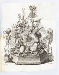
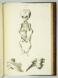
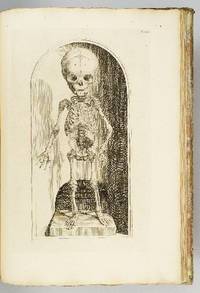
In-8 (mm. 246x191), 2 volumi, p. pelle bazana coeva, dorso a cordoni (restaur.) con decorazioni e titolo oro, tagli rossi, contenenti una straordinaria raccolta completa di 44 opere illustrate (trattati ed epistole, unitamente a lettere in risposta al Ruysch di altri autori), ciascuna con proprio frontesp. e paginazione, e con date di stampa anche precedenti al 1737. Questa edizione è introdotta da: una bellissima antiporta figurata inc. in rame (datata 1720) - un frontespizio con titolo in rosso e nero e vignetta xilografata - elenco delle opere contenute nei due voll. - ritratto dell’Autore in medaglione, inc. in rame e ripieg. Seguono quindi le opere. La collezione è così composta: 1) - "Historia vitae et meritorum Frederici Ruysch" Auctore Ioanne Friderico Schreibero (1732), pp. (8),80. 2) - "Dilucidatio valvularum in vasis lymphaticis, et lacteis" .. Accesserunt quaedam observationes anatomicae rariores (1732), pp. (8),28, con 9 tavole di cui 3 f.t. 3) - "Observationum anatomico-chirurgicarum centuria" (1737). // Segue, con proprio frontespizio: "Catalogus rariorum, quae in Museo Ruyschiano asservantur" (1721), pp. (12),100, con 84 figg. in 41 tavv; 188 (numeraz. continua), con 9 figg. in 5 tavv. 4) - "Adversariorum anatomico-medico-chirurgicorum decas prima".. Cui adjuncta est "Michaelis Ernesti Etmulleri Epistola problematica ad V.C. Fred. Ruyschium De ovario novo" (1729), in 2 parti, ciascuna con proprio frontespizio, pp. (2),34,(2), con 2 tavv.; pp. 18 (la p. 17/18 è stata anteposta nella prima parte). 5) - "Adversariorum anatomico-medico-chirurgicorum decas secunda". (1736), pp. (4),46,(2), con 3 tavv. 6) - "Adversariorum anatomico-medico-chirurgicorum decas tertia". (1723), pp. (4),36,(2), con 3 tavv. 7) - "Johannis Gaubii Epistola problematica, prima (- secunda, tertia).. De pilis, pinguedine, septoque scroti; nec non de papillis piramidalibus; ut & de corpore reticulari, sub cuticula sito, &c." (1724), pp. 32, con 3 tavv. 8) - "Epistola anatomica, problematica quinta", auctore Gerardo Frentz,.. "De vasis sanguiferis periostii tibiæ, ut & viis, per quas vesicula fellea sarcinam acquirit" (1725), pp. 10,(2), con 3 tavv. 9) - "Epistola anatomica, problematica quarta", ad virum clarissimum Fredericum Ruyschium.., "De glandulis, fibris, cellulisque lienalibus, &c. Auctore Joh. Jac. Campdomerco" (1725), pp. 10,(2). 10) - "Epistola anatomica, problematica sexta". Authore Johanne Henrico Graetz.. "De arteria & vena bronchiali, nec non de polypis bronchiorum ejectis, venae & arteriae pulmonalis ramos mentientibus" (1731), pp. 11, con 1 tav. 11) - "Epistola anatomica, problematica septima". Authore Johanne Henrico Grætz,.. "De pia matre, ejusque processibus" (1724), pp. 8, con 1 tav. 12) - "Epistola anatomica, problematica octava". Authore Johanne Henrico Grætz.. "De structura nasi cartilaginea, vasis sanguiferis arteriosis membranæ & cavitatis tympani & ossiculorum auditus eorumque periostio" (1724), pp. 11, con 1 tav. f.t. 13) - "Epistola anatomica, problematica nona". Authore Andrea Ottomaro Goelicke.. "De cursu arteriarum per piam matrem cerebrum involventem, de tertia cerebri meninge, de arteriis membranarum cavitates ossis frontis supra narium radices & eas sub sella equina investientium, nec non de vasis arteriosis novis hepatis & diaphragmatis" (1724), pp. 11, con 1 tav. 14) - "Epistola anatomica, problematica decima". Auctore Bartholomæo Keerwolff.. "De auricularum cordis, earumque fibrarum motricum structura" (1725), pp. 8, con 1 tav. 15) - "Epistola anatomica, problematica, undecima". Authore Joh. Christian. Wolf, Anhaltin.. "De intestinorum tunicis, glandulis, &c." (1721), pp. 10,(2), con 1 tav. 16) - "Epistola anatomica, problematica duodecima". Authore Mich. Ernesto Ettmullero.. "De cerebri corticali substantia, &c." (1721), pp. 28,(4), con 3 tavv. 17) - "Epistola anatomica, problematica tertia & decima". Authore Christiano Wedelio.. "De oculorum tunicis" (1737), pp. 32, con 1 tav. 18) - "Epistola anatomica, problematica, quarta & decima". Authore Mauritio à Reverhorst. "De nova artuum decurtandorum Methodo" (1732), pp. 18,(2), con 2 tavv. 19) - "Epistola anatomica, problematica, quinta & decima". Auctore Alberto Henrico Grætz.. "De vasorum sanguineorum extremitatibus, placentæ uterinæ, mammarumque structura, &c." (1722), pp. 11,(3), con 1 tav. 20) - "Epistola anatomica, problematca decima & sexta". Authore M. Abrahamo Vatero.. "De viis absconditis pulmonum, quibus aër, respirando receptus, in sanguinem penetrat; nec non de vasorum secretoriorum structura mechanica, & de fibrillarum nervearum in cerebro principiis" (1727), pp. 10. 21) - "Frederici Ruyschii .. Thesaurus animalium primus" (1725), pp. (28),20, con 7 tavv. 22) - "Frederici Ruyschii .. Thesaurus anatomicus primus" (1721), pp. 36, con 4 tavv. 23) - "Thesaurus anatomicus secundus" (1722), pp. 46, con 6 tavv. 24) - "Thesaurus anatomicus tertius" (1724), pp. 36, con 4 tavv. 25) - "Thesaurus anatomicus quartus" (1724), pp. 28, con 3 tavv. 26) - "Thesaurus anatomicus quintus" (1725), pp. 28, con 3 tavv. 27) - "Thesaurus anatomicus sextus" (1724), pp. 56, con 7 tavv. 28) - "Thesaurus anatomicus septimus" (1726), pp. 22, con 4 tavv. 29) - "Thesaurus anatomicus octavus" (1727), pp. 32, con 3 tavv. 30) - "Thesaurus anatomicus nonus" (1726), pp. 43, con 5 tavv. 31) - "Frederici Ruyschii .. Thesaurus magnus & regius qui est decimus thesaurorum anatomicorum". In quo præcipuæ corporis humani partes, ceu in statu vivo, nitidissime præparatæ, reservantur (1729), pp. (8),42, con 3 tavv. 32) - ".. Curæ posteriores, seu Thesaurus anatomicus omnium præcedentium maximus" (1724), pp. (10),31, con 3 tavv. 33) - ".. Curae renovatae, seu, Thesaurus anatomicus, post Curas posteriores, novus" (1728), pp. (10),22, con 3 tavv. 34) - "Frederici Ruyschii Responsio ad Godefridi Bidloi libellum", quem vindicias inscripsit (1721), pp. 44. 35) - "Joannis Jacobi Rau Responsio ad qualemcunque defensionem Frederici Ruyschii, quam haud ita pridem edidit, pro septo scroti". In qua hujus litis anatomicæ detegitur origo: et septum scroti, ab ipso descriptum & delineatum, fictitium esse, clare demonstratur. Adjuncta est hujus calci Auctoris Epistola (1737), pp. 16,(2), con 2 tavv. 35 bis) - "Joannis Jacobi Rau Epistola de inventoribus septi scroti, ad virum clarissimum Fredericum Ruyschium", (1737), pp. 6. 36) - "Io. Christoph. Bohlii .. Dissertatio epistolica". Ad virum clarissimum, Fredericum Ruyschium .. "De usu nouarum cavæ propaginum in systemate chylopoeo, ut & de corticis cerebri textura" (1727), pp. 13. 37) - "Frederici Ruyschii .. Responsio, ad Dissertationem epistolicam, viri doctissimi, Joh. Christoph. Bohlii .. quam, de usu novarum cavæ propaginum in systemate chylopoeo, nec non de cortice cerebri, conscripsit" (1727), pp. 12 con 1 fig. nel t. 38) - "Tractatio anatomica, de musculo, in fundo uteri observato", antehac a nemine detecto, cui, accedit depulsionis secundinarum, parturientium feminarum, instructio, authore Frederico Ruyschio .. Ex Belgico in Latinum traducta, a Joanne Christophoro Bohlio (1726), pp. 20, con 1 tav. 39) - "Epistola viri clarissimi Hecqueti", ad D.D. De Ruyschiano uteri musculo (1727), pp. 8. 40) - "Abrahami Vateri. Epistola gratulatoria.. in qua de musculo orbiculari in fundo uteri detecto gratulatur" simulque communicationem eorum quae noviter in anatomia plantarum detexit perquam officiose sibi expetit dubiumque exponit circa lacunas uteri gravidi (1727), pp. 23, con 1 tav. 41) - "Opusculum anatomicum de fabrica glandularum in corpore humano", continens binas epistolas, quarum prior est Hermanni Boerhaave, super hac re, ad Fredericum Ruyschium; altera Frederici Ruyschii ad Hermannum Boerhaave qua priori respondetur (1733), pp. (2),81, con 2 figg. nel t. 42) - "Appendix necessaria, indicis locupletissimi", operum anatomico-medico-chirurgicorum Frederici Ruyschii .. in qua notantur res, quae in Opusculo anatomico, de fabrica glandularum in corpore humano, ab Hermanno Boerhaave et Frederico Ruyschio conscripto, tractantur (1725), pp. 12. 43) - "Frederici Ruyschii.. Operum anatomico-medico-chirurgicorum index locupletissimus, concinnatus ab Ysbrando Gysberto Arlebout" (1725), pp. 105. Rara a trovarsi completa sia di opere che di tavole come nel caso del nostro esemplare. Cfr. Wellcome, IV, pp. 596-601 - Blake 395 - Garrison and Morton, 389 per l’opera ‘Thesaurus anatomicus’: “Ruysch, professor of anatomy at Leyden and Amsterdam, is notable for his method of injecting the vessels. The recipe for the material used by Ruysch has remained a secret. He gave the first description of bronchial blood vessels and vascular plexuses of the heart, demonstrated the valves of the lymphatics, and made a great number of other important discoveries in anatomy”. Leggerm. corto del marg. super.; con uniformi arross. più o meno pesanti, ma complessivam. discreto esemplare.
In-4 (mm. 322x219), p. pergamena coeva (picc. macchie al piatto poster.), dorso a cordoni (ben restaur.) con titolo ms., 18 cc.nn., 340 pp.num., con pregevole frontespizio figurato entro cornice architettonica e ornato da testatine, grandi capilettera figur. a vignetta e finalini, inc. su legno; l’opera è molto ben illustrata da 1 ritratto dell'A. e da 46 superbe tavole anatomiche, inc. in rame, comprese nella numerazione delle pagine. Il colophon porta la data ‘1588’. "Prima edizione in latino". Cfr. Castiglioni, p. 430: "Giovanni Valverde, soprannominato "Huescanus o ab Huesca" dal nome della sua patria Huesca - nel regno di Leòn in Spagna - occupa un posto eminente fra gli anatomisti spagnoli del Cinquecento. Nel 1556 pubblicò una prima ediz. spagnola, tradotta in italiano nel 1560 e in latino nel 1589, che ebbe una grandissima diffusione e fu tra i libri più letti e più studiati del Rinascimento". Cfr. anche Choulant “History and bibliography of anatomic illustration”, p. 206: "This is a Latin translation of the complete text by Valverde, as it appeared in the Italian edition. Besides the title-page of the Italian edition, which is slightly elaborated in the Latin edition, there are given the forty-two original plates by Valverde and four myologic plates by an unknown artist and engraver. At the end of the prefaces is inserted on a whole page a bust of Valverde with the monogram of Niccolò Beatrici. The four newly added engravings represent four muscle-manikins in different positions, all beautifully and forcefully engraved" - Graesse,VII,254 - Sallander,9798 - Hirsch,I,129. Con tracce d’uso nel t. e al frontesp.; qualche lieve arross. margin.; ca. 40 carte (incluse le ultime 10) molto ben restaur. per fori di tarlo al marg. bianco. Malgrado questi difetti è certamente un buon esemplare.
Due parti in un volume in folio (22x31,5 cm) di (16)-464-328 pagine, con 41 illustrazioni calcografiche in cartiglio, antiporta incisa, e ritratto di Scarlattini. Titolo dell'antiporta: Dell'huomo figurato e simbolico. Titolo della seconda parte in occhietto: Dell'huomo indiviso, e nel suo tutto considerato. - Ottime condizioni. Qualche pagina un po' brunita, anche per le variabili caratteristiche della carta, ma esemplare molto fresco. Legatura coeva in mezza pergamena, con titolo manoscritto al dorso. Ex libris nobiliare al retro del piatto. Prima edizione, prima tiratura.
1890179459Peoria Illinois: American Manikin Company 1890. The bare bones of anatomy An educational model depicting male anatomy. It was positioned as a cheaper and more user-friendly alternative to Louis Auzoux's papier-mâché manikins themselves an accessible innovation from earlier wax models. The American Manikin represents a crucial step in the widening accessibility and popularization of anatomy and is a precursor to the mass-produced teaching models of the 20th century. The model is double-sided one face depicting muscles nerves tendons and the spinal column and the other showing muscles veins and bones. The latter face has alphabetically labelled hooks onto which illustrated steel attachments can be affixed. When all the plates are in place the model can be "dissected" by removing each layer in sequence eventually revealing the skeleton. Several of the attachments have additional diagrams on the reverse; for example the attachment illustrating the nerves of the brain also features a diagram of the inner ear on the other side. First produced in 1888 the manikin was designed by Elias Smith 1828-1913 who also published the Atlas of Colored Plates Illustrating Features of Anatomy 1889. Smith was an enterprising businessman who held patents for bicycle saddles and dental equipment. The accompanying advertisement booklet makes extensive reference to the American Manikin's superiority over Auzoux's French model. The attachment for the model's hand is illustrated with the company's calling card. The manikin was issued with 40 attachments of which 24 are present. An accompanying Teachers' Handbook was also produced not present. Wooden model approximately 1070 x 407 mm chromolithographic illustrations affixed to both faces one face with 27 metal hooks. With 24 steel attachments with similar illustrations metal stand comprising base and three attachable legs. Housed in original wooden case 1020 x 510 mm chromolithographic anatomical diagrams printed on inner panels instructional leaflet affixed to same. With 62-page advertisement booklet. Case with near-contemporary postage label of Lewis Bohnett 1880-1970 antique-collector and proprietor of "Trader Lew's Relic Museum" California; "fragile" label dated 1939 to same. Lightly worn from use lacking single hook cockling and a few chips to chromolithographic overlays colours bright; superficial wear to case: in very good condition standing firm. unknown
2322Rome, Domenico de Rossi, 1691, un volume grand in folio (49cm x 38cm), relié en demi-basane à coins, tranches rouges, dos orné de filets dorés (reliure début XIXe), 56 feuillets comprenant 16 feuillets de texte (page de titre incluse) et 40 planches gravées (frontispice inclus)