312 résultats
19750005181Arlington MA: Editions Medicina Rara 1975. Limited edition. printed wrappers. Fine/issued without. Folio grey printed wrappers in Fine publisher's slipcase with 22pp. Introductory booklet in wrappers. <br/><br/>Copy 567 of 2800 facsimiles. Woodcuts. " Soemmering was a professor of anatomy and physiology. Dissection was a focus for him. These works originally published in 1806 and 1809 show his sketches of human auditory organs human taste and vocal organs and olfactory organs. Garrison-Morton 1454 1455 1554. Editions Medicina Rara unknown
185729295Edinburgh: W.H. Lizars 3 St. James Square; S. Highley 32 Fleet St. London and W. Curry Junr & Co 9 Upper Sackville St. Dublin 1857. Hardcover book. Good overall. Large remarkable plates illustrating the human body skeleton organs circulatory muscular and nervous and lymphatic systems fetal respiratory eyes internal organs. The plates with tissue guards most hand colored engraved by W.H. Lizars. <br /> <br /> Owners signature on ffep "George Edward Rundle 30th September 1889" plus his bookplate on inside front cover. Rundle c. 1846 - 1906 was born in Hampshire England and received his education at the University College London and Edinburgh University qualifying in 1873. He first practiced at Hillston NSW and subsequently at Tenterfield and in Sydney by 1876. In July 1875 he was listed as enrolled on the list of medical practitioners of Tasmania in the Launceston Examiner 29 Jul 1875. He is listed in a government gazette with many degrees including a Fellow of the Royal College of Surgeons in Edinburgh in 1878. After retiring he became a trustee of the Sydney Museum and a member of the Zoological Society and for some time had been president. He died 16 Oct 1906 at 14 Wylde Street Potts Point. He is buried at Waverley Church of England cemetery. <br /> <br /> OCLC 338701549 cites 1 copy at the Univ. of York. <br /> <br /> Folio 26pp Contents 101 plates 241pp text xxxvi indices complete. Brown leather and cloth covers detached but present spine partially perished. Internally very clean. W.H. Lizars, 3 St. James Square; S. Highley 32 Fleet St. London, and W. Curry Junr & Co, 9 Upper Sackville St. Dublin hardcover
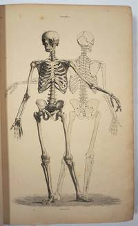
1796015628Cambridge: Printed by J. Burges Printer to the University; And Sold By W. H. Lunn and J. Deighton Cambridge; Messrs. White Fleet-Street and J. Walter Charing-Cross London; And J. Cook Oxford 1796. First Edition First Impression . Cloth Bound Boards. Very Good. 1796 First Edition. Size slim square quarto 72 pages plus 15 pages of plates at the rear. Bound in modern brown cloth Birdsall & Son of Northampton with gilt titles and vignette to the spine. Condition very good corners and spine ends a little rubbed some light spotting and dust soiling at front and rear pages including to the margins of the final plate else the contents are very clean throughout. Contains 15 full page engraved plates at the rear of the book. The plates depict skulls and other bones in the head of various different animals and birds. The title states volume I This was intended as a projected work in two volumes of five fascicules but this first fascicule was the only one ever published. Harwood was a professor of anatomy at Cambridge and an early developer of blood transfusion techniques. OCLC lists only ten copies and no copies sold at auction have been traced. Size: 4to - over 9" - 12" tall <br/> <br/> Printed by J. Burges Printer to the University; And Sold By W. H. Lunn, and J. Deighton Cambridge; Messrs. White, Fleet-Street, hardcover
61852Printed by J. Dixwell No. 148 St. Martin's Lane near Charing Cross. London. 1777. 8vo. pp. 8168 i182-250. Text is continuous despite erratic pagination. Contemporary half calf and marbled boards though now with a later cloth spine occasional foxing more especially at the front ex-libris with stamps and book-label on the front paste-down neat oval stamp on verso of title and in margin of last page overall a very good copy. MAGNUS FALCONAR not in Munk or Plarr was a friend and colleague of William Hewson surgeon anatomist and physiologist; in 1772 Hewson established and ran an anatomy school at 36 Craven Street where Franklin lodged in London now the Benjamin Franklin House museum. In 1998 workmen restoring this building dug up the remains of six children and four adults hidden below the home. The Times reported on 11 February 1998: 'Initial estimates are that the bones are about 200 years old and were buried at the time Franklin was living in the house which was his home from 1757 to 1762 and from 1764 to 1775. Most of the bones show signs of having been dissected sawn or cut. One skull has been drilled with several holes. Paul Knapman the Westminster Coroner said yesterday: "I cannot totally discount the possibility of a crime. There is still a possibility that I may have to hold an inquest." ' The Friends of Benjamin Franklin House noted that the bones were likely placed there by Hewson who lived in the house for two years. They also note that Franklin likely knew what Hewson was doing. Proof was demonstrated by archaeological evidence which showed liquid mercury associated with turtle bones and vermilion colouring associated with dog bones found in the deposit. Hewson had documented experimentation on the lymphatic system using both substances and animals. Hewson died on 1 May 1774 as a result of sepsis contracted whilst dissecting a cadaver. His work was continued after his death by Magnus Falconar who had married Hewson's sister Dorothy in September 1774. Falconar repeated Hewson's experiments on the spleen and thymus and as a result re-published Hewson's work on red blood cells in 1777 together with his corroboration Experimental inquiries: part the third. Containing a description of the red particles of the blood in the human subject and in other animals; with an account of the structure and offices of the lymphatic glands of the Thymus Gland and of the Spleen: being the remaining part of the observations and experiments of the late Mr. William Hewson F. R. S. and Teacher of Anatomy. By Magnus Falconar Surgeon and Teacher of Anatomy. Printed for T. Longman No. 39. Pater-Noster-Row MDCCLXXVII. Samuel Paterson in 1778 published the catalogue of the auction sale of Falconar's anatomical collection - 'Museum falconarianum. A catalogue of the entire and capital museum of anatomical preparations and other subjects of natural history; a great variety of chirurgical anatomical and philosophical instruments; medicaments cabinets preparation-glasses and other effects; of the late Mr. Magnus Falconar surgeon and professor of anatomy deceased: which by order of the adminstrator will be sold by auction by Mr. Paterson at his Great Room No 6. in King-Street Covent-Garden London on Monday the 12th of October 1778 and the nine following evenings to begin precisely at five o'clock. To be viewed on Wednesday the 7th instant and to the time of sale. Catalogues price one shilling may be had at the place of sale; where also may be had Mr. Falconar's synopsis of his course of lectures on anatomy and surgery printed only for the use of his pupils and never before published Price five shillings.' ESTC also records the publication of 'A syllabus of a course of lectures on anatomy on physiology and on the operations and practice of surgery' 1777 Printed by J. Dixwell, No. 148, St. Martin's Lane, near Charing Cross. London. 1777. 8vo. hardcover
174325189Leiden: Joannem & Hermannum Verbeek 1743. From the first edition of one of the greatest of all Anatomical Atlases. Tabula IX which features Albinus and Wandelaar's famous "Muscle Man" a skinned figure whose musculature is visible and defined as viewed in walking profile with the left arm raised and the right foot forward his head is turned slightly away from the viewer. Elephant folio 620 by 475 mm single folio sheet now mounted with the use of none-evasive corner tabs and protected by mylar. Very well preserved fully intact with only the most minor evidence of age. ONE OF THE MAGNIFICENT PLATES FROM "AMONG THE MOST ARTISTICALLY PERFECT OF ANATOMICAL ATLASES." Wandelaar placed his skeletons and musclemen against lush ornamental backgrounds to give them the illusion of vitality using contrasts of mass and light to produce a three-dimensional effect. The most famous plate in the atlas depicts a skeletal figure standing in front of an enormous grazing rhinoceros sketched by Wandelaar from the first living specimen in Europe which had arrived at Amsterdam zoo in 1741" Norman.<br> The plates in this large folio work and in the four supplementary works in large folio with which it is bound are unsurpassed for their cool elegant aesthetic and scientific accuracy. They were drawn and engraved by Jan Wandelaer a pupil of the engravers Jacob Fokema and Guillem van der Gouwen and the painter Gerard de Lairesse who prepared the drawings for Bidloo's atlas. Prior to working for Albinus Wandelaer worked for Friedrik Ruysch. Albinus however provided Wandelaar with the opportunity for the full expression of his talents as a draftsman and engraver. <br> In an attempt to increase the scientific accuracy of anatomical illustration Albinus and Wandelaar devised a new technique of placing nets with square webbing at specified intervals between the artist and the anatomical specimen and copying the images using the grid patterns. Wandelaer placed each figure in a carefully chosen landscape setting and the artistic results are so pleasantly successful that the anatomical figures although composed of many separate parts appear to be actually stepping out of the picture. Joannem & Hermannum Verbeek unknown
174025188Leiden: Joannem & Hermannum Verbeek 1740. From the first edition of one of the greatest of all Anatomical Atlases. Tabula V which features Albinus and Wandelaar's famous "Muscle Man" a skinned figure whose musculature is visible and defined as viewed from behind standing with the left arm raised right arm turned and his weight shifted to the right foot. Elephant folio 620 by 475 mm single folio sheet now mounted with the use of none-evasive corner tabs and protected by mylar. Very well preserved fully intact with only the most minor evidence of age. ONE OF THE MAGNIFICENT PLATES FROM "AMONG THE MOST ARTISTICALLY PERFECT OF ANATOMICAL ATLASES." Wandelaar placed his skeletons and musclemen against lush ornamental backgrounds to give them the illusion of vitality using contrasts of mass and light to produce a three-dimensional effect. The most famous plate in the atlas depicts a skeletal figure standing in front of an enormous grazing rhinoceros sketched by Wandelaar from the first living specimen in Europe which had arrived at Amsterdam zoo in 1741" Norman.<br> The plates in this large folio work and in the four supplementary works in large folio with which it is bound are unsurpassed for their cool elegant aesthetic and scientific accuracy. They were drawn and engraved by Jan Wandelaer a pupil of the engravers Jacob Fokema and Guillem van der Gouwen and the painter Gerard de Lairesse who prepared the drawings for Bidloo's atlas. Prior to working for Albinus Wandelaer worked for Friedrik Ruysch. Albinus however provided Wandelaar with the opportunity for the full expression of his talents as a draftsman and engraver. <br> In an attempt to increase the scientific accuracy of anatomical illustration Albinus and Wandelaar devised a new technique of placing nets with square webbing at specified intervals between the artist and the anatomical specimen and copying the images using the grid patterns. Wandelaer placed each figure in a carefully chosen landscape setting and the artistic results are so pleasantly successful that the anatomical figures although composed of many separate parts appear to be actually stepping out of the picture. Joannem & Hermannum Verbeek unknown
174025187Leiden: Joannem & Hermannum Verbeek 1740. From the first edition of one of the greatest of all Anatomical Atlases. Tabula V which features a human skeleton in a three-quarter view and seen from the rear is portrayed in a walking motion with the left hand raised and extended. Elephant folio ca. 620 by 475 mm single folio sheet now mounted with the use of non-evasive corner tabs and protected by mylar. Very well preserved fully intact with only the most minor evidence of age. ONE OF THE MAGNIFICENT PLATES FROM "AMONG THE MOST ARTISTICALLY PERFECT OF ANATOMICAL ATLASES." Wandelaar placed his skeletons and musclemen against lush ornamental backgrounds to give them the illusion of vitality using contrasts of mass and light to produce a three-dimensional effect. The most famous plate in the atlas depicts a skeletal figure standing in front of an enormous grazing rhinoceros sketched by Wandelaar from the first living specimen in Europe which had arrived at Amsterdam Zoo in 1741" Norman.<br> The plates in this large folio work and in the four supplementary works in large folio with which it is bound are unsurpassed for their cool elegant aesthetic and scientific accuracy. They were drawn and engraved by Jan Wandelaer a pupil of the engravers Jacob Fokema and Guillem van der Gouwen and the painter Gerard de Lairesse who prepared the drawings for Bidloo's atlas. Prior to working for Albinus Wandelaer worked for Friedrik Ruysch. Albinus however provided Wandelaar with the opportunity for the full expression of his talents as a draftsman and engraver. <br> In an attempt to increase the scientific accuracy of anatomical illustration Albinus and Wandelaar devised a new technique of placing nets with square webbing at specified intervals between the artist and the anatomical specimen and copying the images using the grid patterns. Wandelaer placed each figure in a carefully chosen landscape setting and the artistic results are so pleasantly successful that the anatomical figures although composed of many separate parts appear to be actually stepping out of the picture. Joannem & Hermannum Verbeek unknown
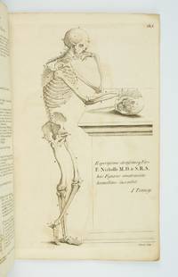
1770ST20219London: Printed for Robert Sayer ca. 1770. 380 x 242 mm. 15 x 9 1/2". 4 leaves of text followed by plates. <br/> Original gray paper wrappers. WITH EIGHT FINE ENGRAVED PLATES of the male body in various poses three depicting the skeleton and five the musculature. Russell 816; Wellcome V p. 273; ESTC N51161. See also Russell "John Tinney's Compendium Anatomicum and its publishers." Wrapper a bit soiled and foxed corners torn with two snags two-inch and half-inch to lower cover most of spine chipped away but the stitching holding the book firmly together; mild offsetting from engravings and a few spots of foxing but a really excellent copy internally the leaves fresh and clean and with very good impressions of the engravings.<br/> <br/> This rare collection of striking anatomical engravings is wonderfully preserved in its original unrestored wrappers. First published in 1743 as "Compendium Anatomicum" the work's plates are adapted from the famous anatomies of Vesalius and Cowper. It was intended as a reference work for artists but as its name suggests it was published with a wider audience in mind. As Tinney humbly tells the reader in the subtitle this is "a work not only very useful but absolutely necessary to painters statuaries and all professors of drawing and design as well as a proper introduction to the study of anatomy for the use of young surgeons" not to mention an "instructive furniture for the studies and libraries of the curious." The work remained in print for a full century though because it was a frequently used book few copies have come down to us in collectible condition. John Tinney ca. 1706-61 was an engraver and print seller who dealt in a wide variety of material being particularly known for maps and satirical prints as well as the present work. Our edition while undated almost certainly dates to the period between 1762-70. The first posthumous edition of "A Compendious Treatise" was released in 1762 by Robert Sayer a map and print seller who was an associate of Tinney's during his lifetime and who likely purchased his stock of plates from his widow following his 1761 death. In 1770 Sayer began a partnership with John Bennett after which the firm was known as "Sayer and Bennett." Since the present edition was published under Sayer's name alone it no doubt comes from the period following the 1762 edition but before the partnership. The present copy has made it through the centuries in remarkably good condition given its flimsy binding and the plates inside with their cheerfully macabre figures remain quite fresh and pleasing. Printed for Robert Sayer unknown
1332007295.Gpaperback. Good. Access codes and supplements are not guaranteed with used items. May be an ex-library book. paperback
1332776078.Gpaperback. Good. Access codes and supplements are not guaranteed with used items. May be an ex-library book. paperback
1920192341920. Original Vintage Photograph from June 1920 of a group of young scientists including a young woman doing a dissection of a human skeletal cadaver. Black and white silver gelatin photograph. 7" x 5" photograph on 8" x 10" board. Captioned "June 1920" on verso. It is likely that this coeducational group are medical students with one older man in glasses seeming to be their instructor. In the background we can see a variety of anatomical drawings as well as bones preserved in liquid in a glass case. The students wear white lab coats except for the female student who is wearing a white gown. While women had entered the nursing profession in large numbers during the 19th century there were still very few female doctors in 1920 making this photo a depiction of a rather uncommon scene. There is a horizontal crack along the upper part of the board which is holding but delicate. The photo itself has a very small closed tear and a small blue smudge in the bottom left corner but is in very good condition overall. unknown
19952081002109000277Japanese Society of Anatomy 1995. Soft Cover. Fine. Number of pages: 387p Size: 31cm Japanese Society of Anatomy paperback