5 848 résultats
51141Paris Chez Jacques Vincent 1750 in 12 (15,5x9,5) 1 volume reliure pleine basane fauve de l'époque, dos à nerfs orné, tranches teintées rouge, 3 feuillets préliminaires non chiffrés, 398 pages. Pierre Chirac, Conques 1648 - Marly-le-Roi 1732, médecin français, premier médecin de Louis XV en 1730. Tome second seul. Bel exemplaire
1830D7399Italy c. 1830. Hardcover. Very Good. Modern half morocco and marbled boards gilt-stamped lettering and ornament on spine; 430 x 560 mm; contains 26 anatomical drawings in red and grey chalk most of them full-page but also including double-page spreads tipped onto stubs. Spine tips and corners gently bumped; some faint dampstaining; a few leaves with repaired tears. Images of skeletal and muscular representations of arms legs feet torsos and heads. <br/><br/> hardcover
201209216Paris, Librairie j-b baillière et fils , 1903 ; in-8, 300 pp., br.
1910123390Paris, Plon-Nourrit et Cie 1910 2 volumes. In-folio 37,5 x 28,5 cm. En feuillets, sous chemise éditeur grise demi-toile rouge à lacets, dos muet, auteur & titre en noir sur le premier plat, XV-270 pp, table des matières - VIII pp. & 110 planches volantes contenant 300 figures dessinées par l’auteur, table des planches. Ensemble en bon état.
1983107102Paris, Maloine 1983 3 volumes. In-4 27,5 x 20,5 cm. Reliures éditeur toile grise, titre bordeaux sur les dos et les pr emiers plats illustrés d’une vignettte polychrome contrecollée, 1843 pp. en pagination continue, plus de 1000 illustrations. Ouvrages en très bon état.
Hardcover in-4° (22 x 30 cm), 202 pages, nombreuses planches en couleurs, cartonnage de l"editeur sous jaquette et emboitage.- Bel exemplaire [QU-4]
201315784, Jena verlag von gustav fischer , 1921 ; in-8, cartonnage de l'éditeur. Les 2 volumes. Band 1 : allgemeine atiologie allgemeine pathologische anatomie band 2spezielle pathologische anatomie en 2 volumes.
201309023Paris, Librairie j-b bailliere et fils , 1954 ; in-8, 306 pp., br. Tome 2.
201506388Monaco, Editions du rocher, 1999 ; in-8, 244 pp., br.
174325189Leiden: Joannem & Hermannum Verbeek 1743. From the first edition of one of the greatest of all Anatomical Atlases. Tabula IX which features Albinus and Wandelaar's famous "Muscle Man" a skinned figure whose musculature is visible and defined as viewed in walking profile with the left arm raised and the right foot forward his head is turned slightly away from the viewer. Elephant folio 620 by 475 mm single folio sheet now mounted with the use of none-evasive corner tabs and protected by mylar. Very well preserved fully intact with only the most minor evidence of age. ONE OF THE MAGNIFICENT PLATES FROM "AMONG THE MOST ARTISTICALLY PERFECT OF ANATOMICAL ATLASES." Wandelaar placed his skeletons and musclemen against lush ornamental backgrounds to give them the illusion of vitality using contrasts of mass and light to produce a three-dimensional effect. The most famous plate in the atlas depicts a skeletal figure standing in front of an enormous grazing rhinoceros sketched by Wandelaar from the first living specimen in Europe which had arrived at Amsterdam zoo in 1741" Norman.<br> The plates in this large folio work and in the four supplementary works in large folio with which it is bound are unsurpassed for their cool elegant aesthetic and scientific accuracy. They were drawn and engraved by Jan Wandelaer a pupil of the engravers Jacob Fokema and Guillem van der Gouwen and the painter Gerard de Lairesse who prepared the drawings for Bidloo's atlas. Prior to working for Albinus Wandelaer worked for Friedrik Ruysch. Albinus however provided Wandelaar with the opportunity for the full expression of his talents as a draftsman and engraver. <br> In an attempt to increase the scientific accuracy of anatomical illustration Albinus and Wandelaar devised a new technique of placing nets with square webbing at specified intervals between the artist and the anatomical specimen and copying the images using the grid patterns. Wandelaer placed each figure in a carefully chosen landscape setting and the artistic results are so pleasantly successful that the anatomical figures although composed of many separate parts appear to be actually stepping out of the picture. Joannem & Hermannum Verbeek unknown
174025188Leiden: Joannem & Hermannum Verbeek 1740. From the first edition of one of the greatest of all Anatomical Atlases. Tabula V which features Albinus and Wandelaar's famous "Muscle Man" a skinned figure whose musculature is visible and defined as viewed from behind standing with the left arm raised right arm turned and his weight shifted to the right foot. Elephant folio 620 by 475 mm single folio sheet now mounted with the use of none-evasive corner tabs and protected by mylar. Very well preserved fully intact with only the most minor evidence of age. ONE OF THE MAGNIFICENT PLATES FROM "AMONG THE MOST ARTISTICALLY PERFECT OF ANATOMICAL ATLASES." Wandelaar placed his skeletons and musclemen against lush ornamental backgrounds to give them the illusion of vitality using contrasts of mass and light to produce a three-dimensional effect. The most famous plate in the atlas depicts a skeletal figure standing in front of an enormous grazing rhinoceros sketched by Wandelaar from the first living specimen in Europe which had arrived at Amsterdam zoo in 1741" Norman.<br> The plates in this large folio work and in the four supplementary works in large folio with which it is bound are unsurpassed for their cool elegant aesthetic and scientific accuracy. They were drawn and engraved by Jan Wandelaer a pupil of the engravers Jacob Fokema and Guillem van der Gouwen and the painter Gerard de Lairesse who prepared the drawings for Bidloo's atlas. Prior to working for Albinus Wandelaer worked for Friedrik Ruysch. Albinus however provided Wandelaar with the opportunity for the full expression of his talents as a draftsman and engraver. <br> In an attempt to increase the scientific accuracy of anatomical illustration Albinus and Wandelaar devised a new technique of placing nets with square webbing at specified intervals between the artist and the anatomical specimen and copying the images using the grid patterns. Wandelaer placed each figure in a carefully chosen landscape setting and the artistic results are so pleasantly successful that the anatomical figures although composed of many separate parts appear to be actually stepping out of the picture. Joannem & Hermannum Verbeek unknown
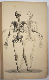
174025187Leiden: Joannem & Hermannum Verbeek 1740. From the first edition of one of the greatest of all Anatomical Atlases. Tabula V which features a human skeleton in a three-quarter view and seen from the rear is portrayed in a walking motion with the left hand raised and extended. Elephant folio ca. 620 by 475 mm single folio sheet now mounted with the use of non-evasive corner tabs and protected by mylar. Very well preserved fully intact with only the most minor evidence of age. ONE OF THE MAGNIFICENT PLATES FROM "AMONG THE MOST ARTISTICALLY PERFECT OF ANATOMICAL ATLASES." Wandelaar placed his skeletons and musclemen against lush ornamental backgrounds to give them the illusion of vitality using contrasts of mass and light to produce a three-dimensional effect. The most famous plate in the atlas depicts a skeletal figure standing in front of an enormous grazing rhinoceros sketched by Wandelaar from the first living specimen in Europe which had arrived at Amsterdam Zoo in 1741" Norman.<br> The plates in this large folio work and in the four supplementary works in large folio with which it is bound are unsurpassed for their cool elegant aesthetic and scientific accuracy. They were drawn and engraved by Jan Wandelaer a pupil of the engravers Jacob Fokema and Guillem van der Gouwen and the painter Gerard de Lairesse who prepared the drawings for Bidloo's atlas. Prior to working for Albinus Wandelaer worked for Friedrik Ruysch. Albinus however provided Wandelaar with the opportunity for the full expression of his talents as a draftsman and engraver. <br> In an attempt to increase the scientific accuracy of anatomical illustration Albinus and Wandelaar devised a new technique of placing nets with square webbing at specified intervals between the artist and the anatomical specimen and copying the images using the grid patterns. Wandelaer placed each figure in a carefully chosen landscape setting and the artistic results are so pleasantly successful that the anatomical figures although composed of many separate parts appear to be actually stepping out of the picture. Joannem & Hermannum Verbeek unknown
185729295Edinburgh: W.H. Lizars 3 St. James Square; S. Highley 32 Fleet St. London and W. Curry Junr & Co 9 Upper Sackville St. Dublin 1857. Hardcover book. Good overall. Large remarkable plates illustrating the human body skeleton organs circulatory muscular and nervous and lymphatic systems fetal respiratory eyes internal organs. The plates with tissue guards most hand colored engraved by W.H. Lizars. <br /> <br /> Owners signature on ffep "George Edward Rundle 30th September 1889" plus his bookplate on inside front cover. Rundle c. 1846 - 1906 was born in Hampshire England and received his education at the University College London and Edinburgh University qualifying in 1873. He first practiced at Hillston NSW and subsequently at Tenterfield and in Sydney by 1876. In July 1875 he was listed as enrolled on the list of medical practitioners of Tasmania in the Launceston Examiner 29 Jul 1875. He is listed in a government gazette with many degrees including a Fellow of the Royal College of Surgeons in Edinburgh in 1878. After retiring he became a trustee of the Sydney Museum and a member of the Zoological Society and for some time had been president. He died 16 Oct 1906 at 14 Wylde Street Potts Point. He is buried at Waverley Church of England cemetery. <br /> <br /> OCLC 338701549 cites 1 copy at the Univ. of York. <br /> <br /> Folio 26pp Contents 101 plates 241pp text xxxvi indices complete. Brown leather and cloth covers detached but present spine partially perished. Internally very clean. W.H. Lizars, 3 St. James Square; S. Highley 32 Fleet St. London, and W. Curry Junr & Co, 9 Upper Sackville St. Dublin hardcover
170434368Leipzig: Thomas Fritsch 1704. First German Edition. Illustrated with an engraved frontispiece of the author and 31 additional full-page engravings depicting various anatomical views. 8vo bound in the original contemporary vellum the spine with calligraphic titling by hand. viii 742 26 Register pp. A very good copy the binding still strong and well preserved the text with the usual mellowing and aging still crisp and very usable. FIRST GERMAN EDITION of this important work on anatomy partnered with the work of Philippi Verheyen Anmerckungen In Die Anatomiam Blancardi. Brieff An den weitberühmten Hrn. Friedrich Ruyschium.<br> 'Verheyen was a prolific writer including several books and manuscripts. His main work published originally in Latin the “Corporis Humani Anatomiae†was translated into multiple languages as here. This book became one of the most used anatomy books of the time.<br> The following excerpt of his book "Corporis Humani Anatomiae Liber Primus" reads : " . QuintusAuricularis quia cum minimum sit auribus expurgandis est aptissimus" translates as".the fifth finger called Auricularis because how small it is is most suitable to clean the ears". Incredibly anatomists at that time called the fifth digit "digitus auricularis".<br> Verheyen is credited with the creation of the eponym the “Achilles tendon†which denominates the common tendon for the gastrocnemius and soleus muscle although at the time he called it the “Chorda Achillisâ€. He also described the kidneys in detail especially the arterial “stars†found on the surface of the kidney which are today known as the “Stars of Verheyen†'. E. A. Miranda 2014 M.T.D. Thomas Fritsch hardcover
1922LFA-126741423Un ouvrage de 650 pages, format 210 x 290 mm, illustré, relié percaline éditeur, publié en 1922, Librairie A. Quillet, bon état
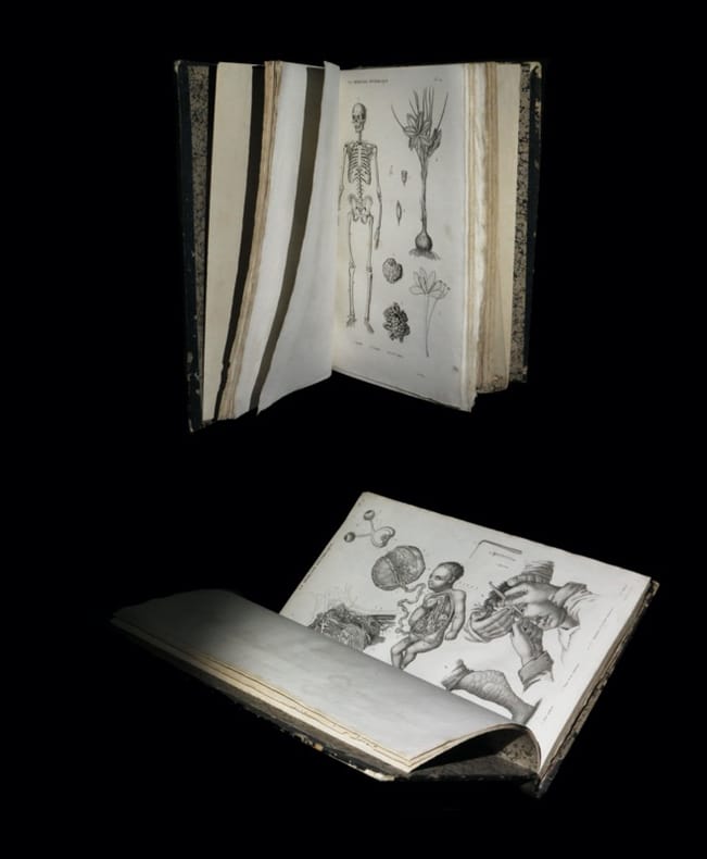
183427522[Page de titre absente] - [Paris, au Bureau de la Médecine pittoresque, 1834 à 1838]. Un vol. au format in-4 étroit (303 x 206 mm) de 1 f. bl., 93 planches, 44 planches et 1 f. bl. Reliure de l'époque de demi-basane glacée marine, dos lisse orné de doubles filets dorés, roulettes dorées, larges fleurons romantiques à froid, titre doré.
1953LFA-126724550Une revue de 32 pages, format 150 x 230 mm, illustrée, brochée, publiée en 1953, bon état
9954de 6 numéros du Progrès Médical, consacrés à Xavier Bichat (1771-1802), célébrissime médecin anatomiste et physiologiste, né à Thoirette alors en Bresse (aujourd'hui dans le Jura), élevé à Poncin dans le Bugey puis à Nantua et Lyon.
1912D2144Paris, Librairie A. Schulz, 1912 ; in-8, 3 pp., broché. Envoi de l'auteur. Bon état.
Paris, Librairie A. Schulz, 1912; in-8, 3 pp., broché. Envoi de l'auteur. Bon état.
1895D0147Bruxelles, Bulletin de la Société Belge de Microscopie, 1895 ; in-8, 11 pp., broché. Tiré à part (extrait). Envoi de l'auteur. Très bon état.
Bruxelles, Bulletin de la Société Belge de Microscopie, 1895; in-8, 11 pp., broché. Tiré à part (extrait). Envoi de l'auteur. Très bon état.
13554P., Albin Michel, 1929, in 8° broché, 131 pages
175244081De l'Imprimerie Royale, Paris 1752. 405 S. mit 17 gefalteten Kupfertafeln, 8°, Interims-Pappband der Zeit, Einband berieben und bestoßen, Papierbezug schadhaft, Stempel auf Titel, mit kleinem Papierrückenschild, innen bis auf vereinzelte Braunflecken sauberes, noch unbeschnittenes Exemplar,
239988Paris, Imprimerie de E. Martinet, 1869 in-8, 48 pp., dérelié.