312 résultats
1796015628Cambridge: Printed by J. Burges Printer to the University; And Sold By W. H. Lunn and J. Deighton Cambridge; Messrs. White Fleet-Street and J. Walter Charing-Cross London; And J. Cook Oxford 1796. First Edition First Impression . Cloth Bound Boards. Very Good. 1796 First Edition. Size slim square quarto 72 pages plus 15 pages of plates at the rear. Bound in modern brown cloth Birdsall & Son of Northampton with gilt titles and vignette to the spine. Condition very good corners and spine ends a little rubbed some light spotting and dust soiling at front and rear pages including to the margins of the final plate else the contents are very clean throughout. Contains 15 full page engraved plates at the rear of the book. The plates depict skulls and other bones in the head of various different animals and birds. The title states volume I This was intended as a projected work in two volumes of five fascicules but this first fascicule was the only one ever published. Harwood was a professor of anatomy at Cambridge and an early developer of blood transfusion techniques. OCLC lists only ten copies and no copies sold at auction have been traced. Size: 4to - over 9" - 12" tall <br/> <br/> Printed by J. Burges Printer to the University; And Sold By W. H. Lunn, and J. Deighton Cambridge; Messrs. White, Fleet-Street, hardcover
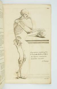
1770ST20219London: Printed for Robert Sayer ca. 1770. 380 x 242 mm. 15 x 9 1/2". 4 leaves of text followed by plates. <br/> Original gray paper wrappers. WITH EIGHT FINE ENGRAVED PLATES of the male body in various poses three depicting the skeleton and five the musculature. Russell 816; Wellcome V p. 273; ESTC N51161. See also Russell "John Tinney's Compendium Anatomicum and its publishers." Wrapper a bit soiled and foxed corners torn with two snags two-inch and half-inch to lower cover most of spine chipped away but the stitching holding the book firmly together; mild offsetting from engravings and a few spots of foxing but a really excellent copy internally the leaves fresh and clean and with very good impressions of the engravings.<br/> <br/> This rare collection of striking anatomical engravings is wonderfully preserved in its original unrestored wrappers. First published in 1743 as "Compendium Anatomicum" the work's plates are adapted from the famous anatomies of Vesalius and Cowper. It was intended as a reference work for artists but as its name suggests it was published with a wider audience in mind. As Tinney humbly tells the reader in the subtitle this is "a work not only very useful but absolutely necessary to painters statuaries and all professors of drawing and design as well as a proper introduction to the study of anatomy for the use of young surgeons" not to mention an "instructive furniture for the studies and libraries of the curious." The work remained in print for a full century though because it was a frequently used book few copies have come down to us in collectible condition. John Tinney ca. 1706-61 was an engraver and print seller who dealt in a wide variety of material being particularly known for maps and satirical prints as well as the present work. Our edition while undated almost certainly dates to the period between 1762-70. The first posthumous edition of "A Compendious Treatise" was released in 1762 by Robert Sayer a map and print seller who was an associate of Tinney's during his lifetime and who likely purchased his stock of plates from his widow following his 1761 death. In 1770 Sayer began a partnership with John Bennett after which the firm was known as "Sayer and Bennett." Since the present edition was published under Sayer's name alone it no doubt comes from the period following the 1762 edition but before the partnership. The present copy has made it through the centuries in remarkably good condition given its flimsy binding and the plates inside with their cheerfully macabre figures remain quite fresh and pleasing. Printed for Robert Sayer unknown
174325189Leiden: Joannem & Hermannum Verbeek 1743. From the first edition of one of the greatest of all Anatomical Atlases. Tabula IX which features Albinus and Wandelaar's famous "Muscle Man" a skinned figure whose musculature is visible and defined as viewed in walking profile with the left arm raised and the right foot forward his head is turned slightly away from the viewer. Elephant folio 620 by 475 mm single folio sheet now mounted with the use of none-evasive corner tabs and protected by mylar. Very well preserved fully intact with only the most minor evidence of age. ONE OF THE MAGNIFICENT PLATES FROM "AMONG THE MOST ARTISTICALLY PERFECT OF ANATOMICAL ATLASES." Wandelaar placed his skeletons and musclemen against lush ornamental backgrounds to give them the illusion of vitality using contrasts of mass and light to produce a three-dimensional effect. The most famous plate in the atlas depicts a skeletal figure standing in front of an enormous grazing rhinoceros sketched by Wandelaar from the first living specimen in Europe which had arrived at Amsterdam zoo in 1741" Norman.<br> The plates in this large folio work and in the four supplementary works in large folio with which it is bound are unsurpassed for their cool elegant aesthetic and scientific accuracy. They were drawn and engraved by Jan Wandelaer a pupil of the engravers Jacob Fokema and Guillem van der Gouwen and the painter Gerard de Lairesse who prepared the drawings for Bidloo's atlas. Prior to working for Albinus Wandelaer worked for Friedrik Ruysch. Albinus however provided Wandelaar with the opportunity for the full expression of his talents as a draftsman and engraver. <br> In an attempt to increase the scientific accuracy of anatomical illustration Albinus and Wandelaar devised a new technique of placing nets with square webbing at specified intervals between the artist and the anatomical specimen and copying the images using the grid patterns. Wandelaer placed each figure in a carefully chosen landscape setting and the artistic results are so pleasantly successful that the anatomical figures although composed of many separate parts appear to be actually stepping out of the picture. Joannem & Hermannum Verbeek unknown
174025188Leiden: Joannem & Hermannum Verbeek 1740. From the first edition of one of the greatest of all Anatomical Atlases. Tabula V which features Albinus and Wandelaar's famous "Muscle Man" a skinned figure whose musculature is visible and defined as viewed from behind standing with the left arm raised right arm turned and his weight shifted to the right foot. Elephant folio 620 by 475 mm single folio sheet now mounted with the use of none-evasive corner tabs and protected by mylar. Very well preserved fully intact with only the most minor evidence of age. ONE OF THE MAGNIFICENT PLATES FROM "AMONG THE MOST ARTISTICALLY PERFECT OF ANATOMICAL ATLASES." Wandelaar placed his skeletons and musclemen against lush ornamental backgrounds to give them the illusion of vitality using contrasts of mass and light to produce a three-dimensional effect. The most famous plate in the atlas depicts a skeletal figure standing in front of an enormous grazing rhinoceros sketched by Wandelaar from the first living specimen in Europe which had arrived at Amsterdam zoo in 1741" Norman.<br> The plates in this large folio work and in the four supplementary works in large folio with which it is bound are unsurpassed for their cool elegant aesthetic and scientific accuracy. They were drawn and engraved by Jan Wandelaer a pupil of the engravers Jacob Fokema and Guillem van der Gouwen and the painter Gerard de Lairesse who prepared the drawings for Bidloo's atlas. Prior to working for Albinus Wandelaer worked for Friedrik Ruysch. Albinus however provided Wandelaar with the opportunity for the full expression of his talents as a draftsman and engraver. <br> In an attempt to increase the scientific accuracy of anatomical illustration Albinus and Wandelaar devised a new technique of placing nets with square webbing at specified intervals between the artist and the anatomical specimen and copying the images using the grid patterns. Wandelaer placed each figure in a carefully chosen landscape setting and the artistic results are so pleasantly successful that the anatomical figures although composed of many separate parts appear to be actually stepping out of the picture. Joannem & Hermannum Verbeek unknown
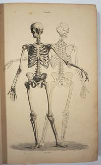
174025187Leiden: Joannem & Hermannum Verbeek 1740. From the first edition of one of the greatest of all Anatomical Atlases. Tabula V which features a human skeleton in a three-quarter view and seen from the rear is portrayed in a walking motion with the left hand raised and extended. Elephant folio ca. 620 by 475 mm single folio sheet now mounted with the use of non-evasive corner tabs and protected by mylar. Very well preserved fully intact with only the most minor evidence of age. ONE OF THE MAGNIFICENT PLATES FROM "AMONG THE MOST ARTISTICALLY PERFECT OF ANATOMICAL ATLASES." Wandelaar placed his skeletons and musclemen against lush ornamental backgrounds to give them the illusion of vitality using contrasts of mass and light to produce a three-dimensional effect. The most famous plate in the atlas depicts a skeletal figure standing in front of an enormous grazing rhinoceros sketched by Wandelaar from the first living specimen in Europe which had arrived at Amsterdam Zoo in 1741" Norman.<br> The plates in this large folio work and in the four supplementary works in large folio with which it is bound are unsurpassed for their cool elegant aesthetic and scientific accuracy. They were drawn and engraved by Jan Wandelaer a pupil of the engravers Jacob Fokema and Guillem van der Gouwen and the painter Gerard de Lairesse who prepared the drawings for Bidloo's atlas. Prior to working for Albinus Wandelaer worked for Friedrik Ruysch. Albinus however provided Wandelaar with the opportunity for the full expression of his talents as a draftsman and engraver. <br> In an attempt to increase the scientific accuracy of anatomical illustration Albinus and Wandelaar devised a new technique of placing nets with square webbing at specified intervals between the artist and the anatomical specimen and copying the images using the grid patterns. Wandelaer placed each figure in a carefully chosen landscape setting and the artistic results are so pleasantly successful that the anatomical figures although composed of many separate parts appear to be actually stepping out of the picture. Joannem & Hermannum Verbeek unknown
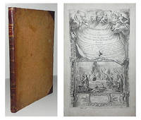
1784000285Etienne Charpentier 1784. Hardcover. See Description. Tall folio 385 mm. The entire book including the text was printed from engraved copper plates 60 plates an illustrated title page plate 29 anatomical plates and 30 calligraphic letterpress plates. Bound in a contemporary or near contemporary binding with calf spine and decorative paste paper boards - rubbed edges shelf worn old ink stain on rear cover. Both the upper portion of the spine and the tail cap are carefully restored. Board corners are neatly repaired. Interior contains an occasional minor spots of marginal foxing; a couple of tiny holes or paper defects are present on the text plate opposite plate 20. The leaves are otherwise generally clean. Francois-Michel Disdier 1708-1781 was a professor of surgery and a drawing master at the Academy of Painting in Paris. His "Tableaux anatomiques" a deluxe anatomy book for physicians was first published in 1758. Many of the plates were done by Crepy and Charpentier some after the style of Vesalius and others following Eustachi. The engraved title page designed by François Bouchet depicts students in an anatomy class studying a cadaver. The whole scene is framed in ornate baroque style. Blake NLM p. 122; Heirs of Hippocrates 894 listing 1778 edition; Hirsch II p. 190. <br/> <br/> Etienne Charpentier hardcover
1830D7399Italy c. 1830. Hardcover. Very Good. Modern half morocco and marbled boards gilt-stamped lettering and ornament on spine; 430 x 560 mm; contains 26 anatomical drawings in red and grey chalk most of them full-page but also including double-page spreads tipped onto stubs. Spine tips and corners gently bumped; some faint dampstaining; a few leaves with repaired tears. Images of skeletal and muscular representations of arms legs feet torsos and heads. <br/><br/> hardcover
185729295Edinburgh: W.H. Lizars 3 St. James Square; S. Highley 32 Fleet St. London and W. Curry Junr & Co 9 Upper Sackville St. Dublin 1857. Hardcover book. Good overall. Large remarkable plates illustrating the human body skeleton organs circulatory muscular and nervous and lymphatic systems fetal respiratory eyes internal organs. The plates with tissue guards most hand colored engraved by W.H. Lizars. <br /> <br /> Owners signature on ffep "George Edward Rundle 30th September 1889" plus his bookplate on inside front cover. Rundle c. 1846 - 1906 was born in Hampshire England and received his education at the University College London and Edinburgh University qualifying in 1873. He first practiced at Hillston NSW and subsequently at Tenterfield and in Sydney by 1876. In July 1875 he was listed as enrolled on the list of medical practitioners of Tasmania in the Launceston Examiner 29 Jul 1875. He is listed in a government gazette with many degrees including a Fellow of the Royal College of Surgeons in Edinburgh in 1878. After retiring he became a trustee of the Sydney Museum and a member of the Zoological Society and for some time had been president. He died 16 Oct 1906 at 14 Wylde Street Potts Point. He is buried at Waverley Church of England cemetery. <br /> <br /> OCLC 338701549 cites 1 copy at the Univ. of York. <br /> <br /> Folio 26pp Contents 101 plates 241pp text xxxvi indices complete. Brown leather and cloth covers detached but present spine partially perished. Internally very clean. W.H. Lizars, 3 St. James Square; S. Highley 32 Fleet St. London, and W. Curry Junr & Co, 9 Upper Sackville St. Dublin hardcover
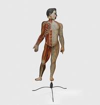
1890179459Peoria Illinois: American Manikin Company 1890. The bare bones of anatomy An educational model depicting male anatomy. It was positioned as a cheaper and more user-friendly alternative to Louis Auzoux's papier-mâché manikins themselves an accessible innovation from earlier wax models. The American Manikin represents a crucial step in the widening accessibility and popularization of anatomy and is a precursor to the mass-produced teaching models of the 20th century. The model is double-sided one face depicting muscles nerves tendons and the spinal column and the other showing muscles veins and bones. The latter face has alphabetically labelled hooks onto which illustrated steel attachments can be affixed. When all the plates are in place the model can be "dissected" by removing each layer in sequence eventually revealing the skeleton. Several of the attachments have additional diagrams on the reverse; for example the attachment illustrating the nerves of the brain also features a diagram of the inner ear on the other side. First produced in 1888 the manikin was designed by Elias Smith 1828-1913 who also published the Atlas of Colored Plates Illustrating Features of Anatomy 1889. Smith was an enterprising businessman who held patents for bicycle saddles and dental equipment. The accompanying advertisement booklet makes extensive reference to the American Manikin's superiority over Auzoux's French model. The attachment for the model's hand is illustrated with the company's calling card. The manikin was issued with 40 attachments of which 24 are present. An accompanying Teachers' Handbook was also produced not present. Wooden model approximately 1070 x 407 mm chromolithographic illustrations affixed to both faces one face with 27 metal hooks. With 24 steel attachments with similar illustrations metal stand comprising base and three attachable legs. Housed in original wooden case 1020 x 510 mm chromolithographic anatomical diagrams printed on inner panels instructional leaflet affixed to same. With 62-page advertisement booklet. Case with near-contemporary postage label of Lewis Bohnett 1880-1970 antique-collector and proprietor of "Trader Lew's Relic Museum" California; "fragile" label dated 1939 to same. Lightly worn from use lacking single hook cockling and a few chips to chromolithographic overlays colours bright; superficial wear to case: in very good condition standing firm. unknown
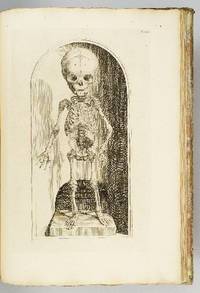
1739ST12883Leyden: Johann Arnold Langerak 1739. First Edition in Latin. 540 x 380 mm. 21 1/4 x 15". 70 leaves of text.Translated by William Dundass. <br/> Original red quarter vellum over marbled boards raised bands UNTRIMMED EDGES. Engraved printer's device on title large decorative initials and tailpieces EXTRA ENGRAVED TITLE AND 114 STRIKING PLATES OF ANATOMICAL FIGURES three folding. Wellcome II 401; Heirs of Hippocrates 468; Choulant-Frank pp. 252-3; Russell 213. ◆Vellum on spine rather worn with three one-and-one-half-inch pieces broken away revealing structure underneath paper boards quite chafed other minor problems externally but an entirely solid unrestored binding. A couple of plates with short closed marginal tears one folding plate with one-inch closed tear into image no loss untrimmed edges a little browned and brittle with isolated small chips occasional minor foxing or insignificant stains three plates lightly browned but still AN UNUSUALLY ATTRACTIVE COPY INTERNALLY with the broadest of margins with especially clean and fresh leaves and with rich impressions of the plates.<br/> <br/> With plates of notable visual impressiveness this was the finest anatomy book in England during the first half of the 18th century; it also was largely a plagiarism borrowing most of its engraved content from a work by the Dutch physician Govert Bidloo published in 1685. Bidloo's work was the first large-scale anatomical atlas to appear after Vesalius' epoch-making "De Humani Corporis Fabrica" and the plates which are highly praised by Norman and Garrison-Morton are characterized by startlingly detailed life-size depictions of the human body both adult and infant with figures flayed to reveal muscles opened to show organs and unfleshed to exhibit bones. According to Choulant-Frank Bidloo's publishers sold 300 impressions of these plates to Cowper probably to recoup some of their money after disappointing sales. Cowper took Bidloo's original 105 plates added nine of his own and produced an English translation of the original Latin text to accompany them. Discussing the original plates produced by Gerard de Lairesse 1641-1711 Norman says that the figures are displayed "in an emotional almost tender manner contrasting the raw dissected parts with the full soft surfaces of uncut flesh placing flayed bound figures in ordinary nightclothes or bedding setting ordinary household objects such as books jars or cabinets in the same scene as cut-up torsos or limbs. His illustrations brought the qualities of Dutch still-life painting into anatomical illustration and gave a new darker spiritual expression to the significance of the act of dissection." When Cowper's version of the atlas first appeared as "The Anatomy of Humane Bodies" in London in 1698 there was also a 1737 Leyden printing in English before our more scholarly Latin edition Bidloo complained to the Royal Society and accused Cowper of plagiarism and fraud resulting in much acrimony and heated pamphleteering between the two physicians. Notwithstanding this scandal Cowper's achievements and discoveries--including the pair of glands that bear his name--were considerable and his text improved significantly upon the original work. Unfortunately as the DNB notes "the notoriety of this case has served to obscure a true appreciation of Cowper and of his many original contributions to anatomical illustration." The atlases of Bidloo and Cowper appear on the market regularly but at 540 x 380 mm. the present copy is distinguished by its size which is significantly larger than what is typically seen with this edition--we have not been able to trace a copy larger than ours from marketplace or institutional records. Johann Arnold Langerak unknown
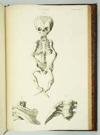
1793ST20586Lugduni Batavorum Leyden: S. & J. Luchtmans 1793 1827 1835. FIRST EDITION A COMPLETE SET. 525 x 350 mm. 20 3/4 x 14". Four volumes. <br/> First two volumes in contemporary cat's paw calf volumes III and IV in contemporary flamed calf raised bands spines gilt in compartments with central fleuron one red and one black morocco label. WITH 206 ENGRAVED ANATOMICAL PLATES all with guards. Front pastedowns with bookplate of Harvard Medical School Library; library shelf markings in white at bottom of spines; bookseller's ticket of Schönhof & Mueller Foreign Books Boston. Bibliotheca Walleriana 8549; Choulant pp. 312-13; Larousse XIX vol. 14 p. 171. Lucas L. Boer Peter L. J. Boek Andries J. van Dam and Roelof-Jan Oostra "History and highlights of the teratological collection in the Museum Anatomicum of Leiden University The Netherlands " American Journal of Medical Genetics Part A Volume 176 no. 3 2018 pp. 618-37.<br /> Shallow chips across the tail of first two volumes covers variably abraded one noticeably so corners somewhat worn partially with boards showing through vague additional signs of wear but the bindings solid and retaining much of their original appeal; plates in first two volumes with faint overall browning either because of paper stock or because of contact with guards a handful of trivial tears but all in all an extremely pleasing set the plates in volume IV very clean and bright and the plates elsewhere and the wide-margined text clean and fresh throughout.<br/> <br/> This is an extremely scarce complete set of a very substantially proportioned anatomical work with more than 200 striking giant folio engravings that emphasize birth defects and that range from clinically cheerless to heartbreaking. A physician and professor of anatomy at Leyden Eduard Sandifort 1742-1814 was commissioned by the University and the city council to publish this account of the school's anatomical collections with a focus on pathological anatomy including especially congenital abnormalities. After the publication of the first two volumes in 1793 the third and fourth were issued following Sandifort's death by his son Gerard 1779-1848 who followed in his father's professorial footsteps in Leyden. The first second and fourth volume are illustrated with copperplate engravings which Larousse tell us make this book "one of the most beautiful works of this genre" though "beautiful" needs to be understood here to mean "convincing" or "compelling" rather than aesthetically appealing. The first volume concludes with nine full-size depictions of skulls; the illustrations in the second and fourth are of a more affecting nature consisting of malformed bodies and body parts damaged or destroyed by disease and birth defects. The engravings of the conjoined twins in these volumes are especially powerful--consisting of poignant even haunting images showing undeserving victims of pronounced congenital defects though some are softened with a touch of the fanciful in their presentation. The thoroughness and accuracy of the work is so great that it remains of scholarly value even today. A recent study by Dutch curators and anatomical scholars Lucas L. Boer Peter L. J. Boek Andries J. van Dam and Roelof-Jan Oostra used "Museum Anatomicum" as a guide to identify and re-diagnose the University's historic teratological collections noting that "many historically made diagnoses could not be improved upon after re-diagnosing the specimens with contemporary dysmorphological knowledge actually confirming that these old collectors were perhaps the first dysmorphologists and can be seen as true pioneers in the field of teratology." Although single volumes and incomplete sets of this work come on the market with some regularity we have been able to trace only five complete sets at auction since 1872. And ours especially fresh and wide-margined in pleasing contemporary calf is a particularly attractive specimen. S. & J. Luchtmans unknown
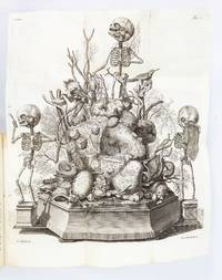
1417ST20185Amsterdam: Joannes Wolters then Jansson-Waesberg 1701-14; 1715-16. FIRST EDITIONS. 224 x 168 mm. 8 3/4 x 6 1/2". 4 p.l. 62 pp. 1 leaf errata; 2 p.l. 98 pp.; 2 p.l. 70 pp.; 1 p.l. 55 pp.; 1 p.l. 54 pp. 2 p.l. 30 pp. 1 leaf 92 pp. 2 leaves 44 pp. 4 leaves 68 pp. 8 leaves 72 pp. 1 leaf errata 12 leaves 78 pp. 10 parts in one volume. <br/> Contemporary stiff vellum smooth spine with ink titling yapp edges. WITH 42 ENGRAVED PLATES as called for by Norman six of these folding. Text in Latin and Dutch. Blake 395; Garrison-Morton 389; Heirs of Hippocrates 627; Jeremy Norman's HistoryofMedicine.com 623; Norman 1875. Vellum lightly soiled front pastedown slightly defective but the original unsophisticated binding in perfect order. Final plate with small hole due to adhesion to facing page minor damage done to image a few other trivial imperfections a couple of negligible marginal tears a faint ink stain across the top inch of text a tiny burn hole but AN UNUSUALLY BRIGHT CLEAN COPY INTERNALLY with all the important plates in exceptionally fine condition.<br/> <br/> Scarce when seen bound from its original 10 parts as here this work describes the memorable "Anatomical Treasure" of Dutch physician and anatomist Frederick Ruysch being illustrated by surrealistic charmingly macabre engravings depicting "fantastic dream-like concoctions constructed of human anatomical parts." History of Medicine Described by Norman as "probably the most original artist in the history of anatomical preparations" Ruysch 1638-1731 "enjoyed making up elaborate three-dimensional emblems of mortality from his specimens" which included fetal skeletons and preserved organs. Heirs to Hippocrates says that "the engraved illustrations deserve special mention for their whimsical almost surrealistic quality: quaintly posed skeletons surrounded by stuffed monsters strange reptiles dried plants and sea creatures." According to Norman "Ruysch's methods allowed him to prepare organs such as the liver and kidneys and keep entire corpses for years. He used a mixture of talc white wax and cinnabar for injecting vessels and an embalming fluid of alcohol made from wine or corn with black pepper added. Using his injection methods Ruysch was the first to demonstrate the occurrence of blood vessels in almost all tissues of the human body thereby destroying the Galenic belief that certain areas of the body had no vascular supply. He was also the first to show that blood vessels display diverse organ-specific patterns." Ruysch's dioramas were displayed in his home museum in Amsterdam which was open to the public; visitors included Tsar Peter the Great of Russia who purchased the collection in 1717 for the foundation of Russia's first public museum the St. Petersburg Kunstkammer. Unfortunately the Russian climate was not kind to the fragile constructions and all deteriorated over time; the dramatic plates here are the best record we have of this unique collection. Joannes Wolters, then Jansson-Waesberg unknown