Weber, Moritz Ignaz (1795-1875)MareMagnum

5 849 résultats
Rappresentazione in scala 1:1 del corpo umano in quattro fogli (660x500 mm ciascuno). Il disegno raffigura la vascolarizzazione venosa ed arteriosa superficiale, evidenziata da bella e vivace coloritura coeva. Inserita nella famosa opera che riproduceva il corpo umano in grandezza naturale "Anatomischer Atlas des Menschlichen Körpers in natürlicher Größe, Lage und Verbindung der Theile in 84 Tafeln und erklärendem" descritta dal Weber. Moritz Ignaz Weber (1795-1875) fu stimato Professore di Anatomia Comparata e Patologica. Spese tutta la sua vita nel campo dell'anatomia e delle scienze associate pubblicando numerosi studi. L’Anatomischer Atlas, che raffigura il corpo umano in 84 tavole litografiche a grandezza naturale, è senza dubbio uno dei suoi lavori più importanti.
First Edition, folio (470 x 290 mm), [5], 6-60pp., 10 engraved plates (4 folding on 2 sheets joined) after A. Fyfe and Thomas Donaldson, plates II and III with short tears not touching the image, plate V a little creased with short closed tears, nineteenth-century half calf, rubbed, covers detached. This was "The first serious study of this subject and the most original anatomical work by the greatest of the Monro dynasty." It contains the first full anatomical description of the sacs between the tendons and bones which Albinus had named the bursae mucosae. They are illustrated on the ten plates "which for explicit clarity and accuracy have not been improved upon." (Heirs of Hippocrates 1011). The plates depict the foot, various joints including the knee and hip, and four of the plates are life-sized representations of the entire arm and leg. Monro secundus' earlier publications were largely polemical, and it was not until he had been teaching for twenty-five years that his three main contributions to medical literature appeared. Monro's Observations on the Structure and Functions of the Nervous System (Edinburgh, 1783), a massive text and atlas on human and comparative neurology, is Monro's greatest work. His Description of All the Bursae Mucosae of the Human Body... was a practical manual for direct use in surgery. Although next to nothing was known of germ life at that time, Monro's acute observation and independent empirical judgement led him to the conclusion that the chief danger of infection in surgery of joints lay in exposure to the air. Garrison-Morton, 399.2; Blake, p.309; Heirs of Hippocrates 1011; Wellcome, IV, p.156. Russell, British anatomy, 613. Taylor, The Monro Collection, M170.
178230054DB2 Bände. Leipzig, Johann Friedrich Junius, 1782. 8°. XXVIII, 886 (recte 884) S; XVI, 811 (recte 809) S., (71) S. Mit 10 gest., gef. Kupfertafeln. Halblederbände aus der Zeit mit 2 verschiedenfarbigen Rückenschildern und reicher Rückenvergoldung.
178230054AB2 Bände. Leipzig, Johann Friedrich Junius, 1782 8°. XXVIII, 886 (recte 884) S.; XVI, 811 (recte 809) S., 71 n.n. S. Mit 10 gestochenen, gefalteten Kupfertafeln. Halblederbände der Zeit mit 2 verschiedenfarbigen Rückenschildern und reicher Rückenvergoldung.
178230054ABLeipzig, Johann Friedrich Junius, 1782. 8°. XXVIII, 886 (recte 884) S.; XVI, 811 (recte 809) S., 71 n.n. S. Mit 10 gestochenen, gefalteten Kupfertafeln. Halblederbände der Zeit mit 2 verschiedenfarbigen Rückenschildern und reicher Rückenvergoldung. 2 Bände.
1796015628Cambridge: Printed by J. Burges Printer to the University; And Sold By W. H. Lunn and J. Deighton Cambridge; Messrs. White Fleet-Street and J. Walter Charing-Cross London; And J. Cook Oxford 1796. First Edition First Impression . Cloth Bound Boards. Very Good. 1796 First Edition. Size slim square quarto 72 pages plus 15 pages of plates at the rear. Bound in modern brown cloth Birdsall & Son of Northampton with gilt titles and vignette to the spine. Condition very good corners and spine ends a little rubbed some light spotting and dust soiling at front and rear pages including to the margins of the final plate else the contents are very clean throughout. Contains 15 full page engraved plates at the rear of the book. The plates depict skulls and other bones in the head of various different animals and birds. The title states volume I This was intended as a projected work in two volumes of five fascicules but this first fascicule was the only one ever published. Harwood was a professor of anatomy at Cambridge and an early developer of blood transfusion techniques. OCLC lists only ten copies and no copies sold at auction have been traced. Size: 4to - over 9" - 12" tall <br/> <br/> Printed by J. Burges Printer to the University; And Sold By W. H. Lunn, and J. Deighton Cambridge; Messrs. White, Fleet-Street, hardcover
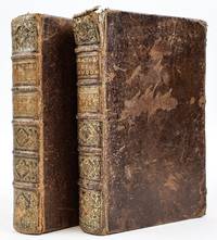
170838094ABLeipzig Thomas Fritschen 1708. 2., deutsche Ausg. 8° 5 Bll., 742 S., 12 Bll., m. gest. Porträt u. 31 Kupfertaf. Pergament. d. Zt. Leicht stockfl. u. wasserrandig, Tit. a. unt. Rand knapp beschnitten, Vors. gestempelt u. m. Besitzvermerken. - Second German edition. Engraved portrait and 31 copperplates. Contemp. vellum. - Slightly foxed and waterstained, title on the foot of page cropped close, endpaper stamped and with ownership entries. 2
174417912Leyde Langerak & Verbeek 1744 In-folio 280 pp et nombreuses figures en 46 planches avec la même au trait en regard, Tranches cirées, re-Relié dans un vélin moderne non vieilli. complet des planches d'après la table. il s'agit d'un commentaire sur les travaux d'Eustacchio ( 1520-1574) de Rome dont les premières planches ont seulement été publiées par Lancisi en 1714 et qui fut un adversaire de Vésale. texte en Latin, première édition.
In-8°. Pagine (8), 716, (2), 24, 13 cc. di tavv. Rilegatura in mezza pelle un po' sciupata e con mancanze al dorso. Stato di conservazione ottimo. Alla fine sono presenti 13 tavole anatomiche ripiegate di eccellente fattura. Rara prima edizione francese di questo celebre testo di anatomia del medico e chirurgo tedesco Lorenz Heister, assai difficile a trovarsi completo di tutte le sue 13 tavole.
3456P., Méquignon, 1825, 2 VOLUMES in 8 DE TEXTE et 1 ATLAS in 4, brochés, couvertures imprimées, (quelques rousseurs, quelques mouillures pâles dans les marges intérieures des derniers feuillets du tome 2, petites et habiles restaurations à l'angle supérieure de la page de couverture, dans la marge extérieure de la page de titre et des feuillets de texte de l'atlas, inversion de deux feuillets de texte à l'atlas), VOlUME 1 (1ère partie) : 27pp., 360pp., VOlUME 2 (2ème partie) : (2), pp. 355/801, (1), ATLAS : 12pp., 13 PLANCHES.
4179Leipzig & Heidelberg, Winter'sche Verlagshandlung, 1861, un volume de texte et un atlas in 8 reliés en demi-basane havane à coins (reliure de l'époque), (déchirure sans manque de papier dans la marge inférieure de la planche 27), Texte : 4pp., 243pp., Atlas : 50 planches lithographiées EN COULEURS
5190Amsterdam, Joannem Janssonium, 1666; un volume in 4 relié en plein veau, dos orné de fers dorés (reliure de l'époque), (restauration ancienne à la partie inférieure du dos, quelques épidermures sur les plats et à un mors, petit travail de vers dans la marge supérieure de 6 feuillets, tache d'encre dans la mage inférieure d'un feuillet, quelques cahiers uniformément roussis), 1 PORTRAIT DE BLASIUS, 1 TITRE GRAVE, (11), 558pp., (8), 52 PLANCHES à pleine page.
18304759DBDüsseldorf, Arnz, )1830-1833). Doppelfolio. Titelblatt, 46 teilweise kolorierte Lithographien. Halbleinenband mit Pappdeckeln. + Wichtig: Für unsere Kunden in der EU erfolgt der Versand alle 14 Tage verzollt ab Deutschland / Postbank-Konto in Deutschland vorhanden +, 4759DB|4759DB_2|4759DB_3 [3 Warenabbildungen]
Rappresentazione in scala 1:1 del corpo umano in quattro fogli (660x500 mm ciascuno). Il disegno raffigura l'apparato muscolo scheletrico anteriore. Inserita nella famosa opera che riproduceva il corpo umano in grandezza naturale "Anatomischer Atlas des Menschlichen Körpers in natürlicher Größe, Lage und Verbindung der Theile in 84 Tafeln und erklärendem" descritta dal Weber. Moritz Ignaz Weber (1795-1875) fu stimato Professore di Anatomia Comparata e Patologica. Spese tutta la sua vita nel campo dell'anatomia e delle scienze associate pubblicando numerosi studi. L’Anatomischer Atlas, che raffigura il corpo umano in 84 tavole litografiche a grandezza naturale, è senza dubbio uno dei suoi lavori più importanti.
17341408797Paris: Guillaume Cavelier 1734. Later Editions. Hardcover. Octavos Four Volumes bound as two. The first book contains a single volume; the second book contains the second volume of Anatomie Chirurgicale in addition to two other standalone works. In Good condition. In full brown leather bindings; spines paneled with elaborate gilt decoration and gilt titling on tan leather labels; gilt roll to board edges. Scuffing and wear to boards with peeling to covers. All edges and fore corners rubbed with sections of exposed board. Chipping to heads and tails of spines; heaviest at head of Vol. I with head band nearly detached. Text block edges show shelf wear and dust soiling. Lightly age toned and very sparsely foxed throughout. Splitting to hinges. Rear pastedown in Vol. I and both pastedowns in Vol. II are separating from the boards. ~2 in. closed tear in Vol. I p. 39. Splitting in gutters of several signatures throughout both volumes due to damage to end bands. Closed tears affect some fold-out plates. Tidemarks affect pages in the rear of both volumes.<br /> <br /> <br> <br> <br /> <br /> CONTENTS: Anatomie Chirurgicale Vol. I xxiv 514 2 pages plus plates i-xxx; Anatomie Chirurgicale Vol. II viii 313 336-352 453-465 366-403 pages plus plates xxxi-xlviii bound together with Observations Anatomiques viii 175 pages plus 8 plates and Six Observations 2 36 2 pages. Shelved in Room A. The first work in this set is the second edition of Flemish surgeon Jean Palfin's Jan Palfijn's very famous treatise on anatomy which was first published in 1726 under the title Anatomie du Corps Humain. This work and its earlier edition were very influential in the medical field for their detailed anatomical illustrations and descriptive actionable advice for surgeons becoming a widespread authoritative medical text throughout Europe and other parts of the world. <br /> <br /> <br> <br> <br /> <br /> The second work bound together with Vol. II of Anatomie Chirurgicale is a French translation of Frederik Ruysch's Observationum Anatomico-Chirurgicarum Centuria. Ruysch was a renowned anatomist and botanist in his own right most well-known for his anatomical museum - the Cabinet of Curiosities - and his improved embalming technique which preserved bodies for longer periods of time in a more visually life-like state.<br /> <br /> <br> <br> <br /> <br /> The final work also bound together with Vol. II of Anatomie Chirurgicale and Observations Anatomiques is a revised edition of Six Observations by Michel Brisseau who is notable for being the first to demonstrate the true nature of cataracts as an increased opacity of the lens of the eye rather than a film that covered the eye which was the prevailing belief at the time. Together these works comprise a collection of highly influential studies treatises and observations on anatomy and surgery by some of the most notable medical practitioners of the late 17th and early 18th centuries. 1408797. Special Collections. Guillaume Cavelier hardcover
18304759DBDüsseldorf, Arnz, )1830-1833). Doppelfolio. Titelblatt, 46 teilweise kolorierte Lithographien. Halbleinenband mit Pappdeckeln.
183346441833 demi-bas.bleue. atlas in-folio de 39pp. et 41 planches en couleurs, Paris J.B. Baillière 1833,
In -8°, pp. 12, cartonato moderno. Paolo Manfredi (1640-1716), lucchese di nascita, visse a Roma dove studiò al Collegio Romano e poi alla Sapienza, dove si sarebbe dedicato alla medicina. Dopo aver studiato per qualche anno le trasfusioni da uomo a uomo e da animale a uomo, ottenne un posto di lettore ordinario di anatomia e chirurgia nel 1668, poco prima della redazione di questo racconto di una dissezione, volume che, nel suo stile, accorda “un approccio strettamente dimostrativo con uno più tradizionalmente retorico-filosofico” (DBI, sub vocem) Paolo Manfredi (1640-1716), born in Lucca, lived in Rome where he was a student at the Collegio Romano and then at La Sapienza University, where he devoted himself to medicine. After having studied some years the blood transfusion (even the animal-to-man one), he obtained a chair as a teacher in anatomy and surgery in 1668, before drafting this dissection tale, a volume that, in Manfredi’s style, is written halfway between demonstration and philosophy (see Dbi, sub vocem)
161419374Spacheri 1614 in-8 plein vélin 2 textes en un volume, reliure janséniste d'époque en plein vélin ivoire parcheminé (jansenist's binding full vellum) in-octavo (22 x 17 cm), dos long (spine without raised band), dos muet (spine without title), plats muets (cover without text), toutes tranches lisses (all smooth edges), orné d'un "Merveilleux" titre gravé sur bois en noir , texte en latin , pagination au feuillet (2 pages) 34 feuillets (68 pages) pour "PINAX MICROCOSMOGRAPHICUS..." et 12 feuillets (24 pages) pour "ELUCIDARIUS TABULIS SYNOPTICIS...", 1615 (S. l.,) : sumptibus S. M. Spacheri (Editeur),
Rappresentazione in scala 1:1 del corpo umano in quattro fogli (660x500 mm ciascuno). Il disegno raffigura l'Rappresentazione in scala 1:1 del corpo umano in quattro fogli (660x500 mm ciascuno). Il disegno raffigura l'apparato muscolo scheletrico posteriore. Inserita nella famosa opera che riproduceva il corpo umano in grandezza naturale "Anatomischer Atlas des Menschlichen Körpers in natürlicher Größe, Lage und Verbindung der Theile in 84 Tafeln und erklärendem" descritta dal Weber. Moritz Ignaz Weber (1795-1875) fu stimato Professore di Anatomia Comparata e Patologica. Spese tutta la sua vita nel campo dell'anatomia e delle scienze associate pubblicando numerosi studi. L’Anatomischer Atlas, che raffigura il corpo umano in 84 tavole litografiche a grandezza naturale, è senza dubbio uno dei suoi lavori più importanti. Inserita nella famosa opera che riproduceva il corpo umano in grandezza naturale "Anatomischer Atlas des Menschlichen Körpers in natürlicher Größe, Lage und Verbindung der Theile in 84 Tafeln und erklärendem" descritta dal Weber. Moritz Ignaz Weber (1795-1875) fu stimato Professore di Anatomia Comparata e Patologica. Spese tutta la sua vita nel campo dell'anatomia e delle scienze associate pubblicando numerosi studi. L’Anatomischer Atlas, che raffigura il corpo umano in 84 tavole litografiche a grandezza naturale, è senza dubbio uno dei suoi lavori più importanti.
624Gottingen, Dieterich, 1805, un volume in 8 relié en demi-basane à coins, dos orné de filets dorés, étiquette rouge, (reliure postérieure), (quelques rousseurs), 16pp., 549pp., (1pp.), (1), 8 PLANCHES
183911062Paris, Furne 1839. Mit 1 gestochenem Porträt, 6 Titelvignetten, 5 grenzkolorierten gestochenen Karten u. 114 handkolorierten gestochenen Tafeln. 4°. Himbeerrote Halblederbände der Zeit mit Rückenvergoldung. Etwas berieben, Ecken u. Kanten etw. beschabt u. bestoßen. Teils etw. gebräunt, einzelne Taf. u. Bl. stärker. Innengelenke teils locker. In Bd. 1 u. 6, S. 545-Schluß vertauscht. - Sehr schöne Ausgabe der Werke des berühmten französischen Naturforschers.
In-4 (mm. 323x215), cartonato rustico coevo, pp.nn. 28 di spiegazione delle 24 tavole anatomiche inc. in rame, ciascuna con numerose figure. Queste popolarissime tavole illustravano l’opera del Vesling “Syntagma anatomicum”, pubblicata per la prima volta nel 1741 e divenuta così popolare da essere ristampata più volte - anche senza testo - come la presente edizione (la prima è del 1709). “On these prints Giovanni Georgi is named as the artist or engraver”, come specifica Choulant, p. 243. Jan Vesling, celebre anatomista tedesco (1599-1649), studiò e svolse la sua attività in Italia. "A Venise il fit des cours particuliers d'anatomie et de botanique, avec un tel succès, que les élèves désertaient les écoles publiques pour venir profiter ses leçons. La République le nomma en 1632 à la première chaire d'anatomie, vacante à l'université de Padoue". Cosi' Biographie Universelle, XLVIII, p. 310. Con timbri di apparten. ma esemplare ben conservato. .
FIRST EDITION OF THE FIRST SCIENTIFIC/GEOMETRICAL STUDY OF HUMAN MOVEMENT. xii, 295 pp plus three folding tables with numerous complex diagrams. Printed on fine wove paper. 8vo. Original wraps. Uncut and unopened. Backstrip mostly gone, else a PRISTINE COPY OF A VERY RARE AND IMPORTANT WORK.
In 8°(mm 205x135). Piena pergamena coeva con titolo manoscritto al dorso, tagli a spruzzo in rosso; pagg. [8], 215, [1], XXVIII [i.e. 26] carte di tav. compreso l'antiporta raffigurante lo studio di anatomia, frontespizio stampato in rosso e nero; iniziali e fregi xil. La tavola XVI è numerata XVI-XVII, la XXII è numerata XXII-XXIII. <BR>Testo latino, su due colonne. Poco comune edizione romana di questo manuale illustrato di anatomia a cura del medico tedesco Johann Adam Kulm (1689-1745); l'edizione originale, è del 1722, poi fu tradotta in latino nel 1731. Si tratta del primo libro occidentale tradotto in giapponese e pubblicato in Giappone (1774). <BR>Bell'esemplare su carta forte, occasionali fioriture. Iscrizione d'appartenenza manoscritta al contropiatto anteriore ("ad usum Josephi Aloysi Ma.... " - non facilmente leggibile).<BR>Valleriana cita altre edizioni; Choulant, pag. 34"in 1722 by imitations of Verheyen's plates by the Danzig physician Johann Adam Kulmus, which ran through many editions and were in part revised and provided with new engravings. They attained a wide circulation. Many other anatomic textbooks of this period had no pictures at all or at least, no complete series, since the aim was to produce books as cheaply as possible for the use of the student.<BR>Uncommon Roman edition of this illustrated manual of anatomy by the German physician Johann Adam Kulm (1689-1745); the original edition, in German, had been published in 1722, then was translated into Latin in 1731; it was the first western book to be translated and published in Japan and Japanese (1774).<BR><BR>