5 849 résultats
in 4° (cm 27x21), pp. 2 b., 568, 2 b. testo; 2 b., 536, 2 b. spiegazione delle tavole; 2 voll. contenenti 340 tavole litografate protette da veline; bella leg. coeva in m/pelle con tit. e fregi in oro ai ds. (abilmente restaurati), piatti in legno foderati di carta marrone glassata, sguardie marmor.; front. allegorico al 1° vol. di tav.; importante opera di anatomia, Brunet II, p. 110; Graesse II, p. 203. lievi fior. dovute alla qualità della carta, e trascurabili tracce di tarlo al margine inf. di poche pagg. ma ottimo esemplare. Ex libris. 593/23
1758839Paris, E. Charpentier, 1758. 60 num. gestochene Blätter (incl. Titel, 29 anatomischen Tafeln und je 1 Bl. Erklärung). Folio. Mod. Pappband mit Rückenschild.
170019096Venedig, Poletti, 1700. 10 Bll., 339 (ohne S. 3 bis 6), LXXXVI S. mit 20 ganzseit. Textkupfern. 1 Kuper-Frontispiz. 8°. Pgmt. der Zeit (etw. gebräunt und fleckig, bestoßen, kl. resturierte Läsuren). [5 Warenabbildungen]
1784000285Etienne Charpentier 1784. Hardcover. See Description. Tall folio 385 mm. The entire book including the text was printed from engraved copper plates 60 plates an illustrated title page plate 29 anatomical plates and 30 calligraphic letterpress plates. Bound in a contemporary or near contemporary binding with calf spine and decorative paste paper boards - rubbed edges shelf worn old ink stain on rear cover. Both the upper portion of the spine and the tail cap are carefully restored. Board corners are neatly repaired. Interior contains an occasional minor spots of marginal foxing; a couple of tiny holes or paper defects are present on the text plate opposite plate 20. The leaves are otherwise generally clean. Francois-Michel Disdier 1708-1781 was a professor of surgery and a drawing master at the Academy of Painting in Paris. His "Tableaux anatomiques" a deluxe anatomy book for physicians was first published in 1758. Many of the plates were done by Crepy and Charpentier some after the style of Vesalius and others following Eustachi. The engraved title page designed by François Bouchet depicts students in an anatomy class studying a cadaver. The whole scene is framed in ornate baroque style. Blake NLM p. 122; Heirs of Hippocrates 894 listing 1778 edition; Hirsch II p. 190. <br/> <br/> Etienne Charpentier hardcover
10445Patavi [Padoue], Paul Frambotti, 1647 ; in-4. Titre gravé-Titre-6ff.- 274pp.-6ff. (table et poèmes dédiés à l'auteur) - 24 planches hors-texte gravées en taille douce (complet). Veau fauve, dos à nerfs orné de petits fers dorés, doubles filets d'encadrement sur les plats ; mouillure pale en pied sur plusieurs feuillets. Reliure restaurée. Petite note biographique manuscrite de 5 lignes, en latin, à propos de Vesling, sur la première garde. Provenance : ex-libris manuscrit sur le titre typographique : Jacques Senné, médecin de Montpellier. Un billet manuscrit signé du même Senné nous apprend qu'il résidait à Marennes (Charente Maritime).
In Fiorenza, nella Stamperia di Zanobi Pignoni, 1643, volume unico, in-4, legatura settecentesca che riutilizza due pergamene più antiche (probabilmente smontate da rilegature di libri di formato inferiore), con dorso rinforzato in carta decorata antica e titolo calligrafato su tassello in carta, pp. 610, [2]. Frontespizio stampato in rosso e nero con stemma mediceo, 23 incisioni xilografiche nel testo (fra cui forme geometriche, la pianta di un passaggio segreto, mobili a doppio fondo, teschi e ossa umane) e una tavola allegorica in rame fuori testo (che quasi mai si trova) raffigurante un liuto senza una corda su cui si poggia una cicala e con motto latino "Ut suppleat" - simbologie per la stonatura delle perversioni del genere umano, sostenuta però dall'intelletto personificato dall'apollinea cicala -. Rara opera seicentesca sul diritto criminale scritta da Antonio Maria Cospi, giudice in varie sedi dello Stato toscano fra la fine del Cinquecento e l'inizio del Seicento, ed edita postuma dal nipote Ottaviano. Si tratta di un testo di pratica giudiziaria che tenta di organizzare i ricordi e le conoscenze professionali dell'autore. Si spazia infatti dalla trattazione scientifica alla spicciola quotidianità del criminalista, passando per la superstizione e l'aneddotica. Il Cospi si sofferma nella prima parte sulla deontologia del giudice, indicando la lussuria e quindi - in una visione fortemente antifemminile - la donna come "ianua diabuli". Nella seconda parte tratta gli ambiti della criminalità, soprattutto quelli fra reato e peccato, quali la magia, la divinazione, l'eresia, la stregoneria. Nella terza, oltre che su negromanzia, avvelenamenti, stupri, aborti, ladri, zingari e bari (alle carte o ai dadi), si sofferma a lungo sull'importanza della ricognizione del cadavere e del luogo del suo ritrovamento. Ci troviamo di fronte a un quasi manuale per "la scena del crimine", con tanto di descrizioni dettagliate e illustrazioni. L'autore sostiene proprio che il giudice necessiti di conoscenze scientifiche, anatomiche, chimiche e di disegno, oltre a quelle prettamente giuridiche e morali, per poter analizzare al meglio e mettere a verbale ogni caso in modo critico. Altro concetto innovativo e fondamentale è quello della testimonianza, intesa come interrogatorio al fine di trarre indizi dall'esame del reo e vista come unica via laddove la scienza non può arrivare. Dalle bibliografie a nostra disposizione (Piantanida/Diotallevi/Livraghi 1948, Michel 1970 e la rivista "Le Carte e la Storia" edita da Il Mulino 2002) questa del 1643 risulterebbe come prima edizione, anche se, probabilmente in tempi successivi, ne sarebbe stato censito un unico esemplare, alla Biblioteca Nazionale di Firenze, datato invece 1638. Più probabilmente però, secondo la nostra interpretazione, si tratterebbe di un errore tipografico sulla data di quest'ultimo esemplare: MDCXXXVIII (1638) invece di MDCXXXXIII (1643). La carta Oo presenta uno strappo con perdita di qualche carattere; minuto camminamento di tarlo alle 6 carte da Vv3 a Xx4, solamente marginale; alcuni fascicoli leggermente bruniti per la fattura della carta. Comunque un bell'esemplare.
ORD-2754Recueillies et publiées sous ses yeux par C. DUMÉRIL, chef des travaux anatomiques de l'École de Médecine de Paris. Paris. Baudouin. An VIII - An XIV (1800-1805). 5 tomes en 5 volumes in-8 (140 x 225mm) dos lisses basane brune, frises or, pièces de titre maroquin rouge, de tomaison, maroquin vert, tome 1: 2ff.n.ch., XXXI, 521, (1) pages, 9 tableaux sur 6 planches dépliantes; tome 2: 2ff.n.ch., XVI, 697, (3) pages; tome 3: 2ff.n.ch., XXVIII (les 8 premières sont à la fin de l'ouvrage), 558, (2) pages; tome 4: 2ff.n.ch., XII, 539, (1) pages; tome 5: 2ff.n.ch., VII, 368 pages, 52 planches (la 52 est placée après la planche 18). Défauts d'usage, rousseurs sur le texte qui ont épargné les planches. Plutôt bon exemplaire, non rogné, bien complet de ses planches, tableaux et errata. Edition originale.
In -4°, pp. 15, 1 tav.; legatura in carta decorata. L’autore era Membro dell’Accademia fisicomatematica di Giusto Ciampini, piccola accademia che rientrava nella protezione di Cristina di Svezia, della quale fu anche segretario. L’opera si presenta in forma di lettera indirizzata a Francesco Redi: nel trattato si discute dell’origine di una grave deformità alla nascita, in una dimensione ricollegabile più al campo, fortemente dibattuto all’epoca, della teoria della generazione. Nota di possesso manoscritta: “Francisci Mariae Campioni”, ecclesiastico ed erudito dell’epoca. Edizione originale. The author was a member of Accademia fisicomatematica di Giusto Ciampini, a small roman academy put under the protection of Christina Queen of Sweden, academy that he was also secretary of. This work is posed as a letter to Francesco Redi: the heavy anatomical aberration of a newborn is discussed, in a optic strictly linked to the field, widely discussed at time, of the generation theory. An ownership inscription under the title page: “Francisci Mariae Campioni”, who was a contemporary ecclesiastic and scholar. Original edition.
In-4°; pp. (12), 588, (4) e in antiporta ritratto dell’autore inciso all’acquaforte nel 1716 da Donato Creti. Opera filosofica circa l’anima sensitiva e l’anima conoscitiva, che prende le mosse dal trattato del 1672 del gesuita matematico e fisico Ignace-Gaston Pardies. Sbaragli si inserisce nella controversia circa l’anima degli animali, che fu affrontata anche nel sistema meccanicistico cartesiano (il quale in verità per gli animali parlava di res cogitans, e non di anima). L’opera è strutturata in tre disputationes: An Bruta sint instar Automatum conformata, An auctoritates Sacrae scripturae sit in physicis attendenda et quantum sit illi tribuendum, An animae brutorum educantur de potentia materiae. Legatura in piena pergamena con titolo in oro al dorso. Blake p. 403. Giovanni Girolamo Sbaraglia. (Bologna 1641 - ivi 1710) si laureò in Filosofia e Medicina a Bologna, e vi insegnò Logica e Medicina Teorica. bologna filosofia philosophy controversy souls beasts anima cartesio medicina anatomia anatomy pardies descartes automata gassendi
17969532Belitz u. Braun, Berlin 1796. IV, 176 u. II, 192 S. mit 5 handkolorierten gestochenen Tafeln nach P. Mügge von C.C. Glaßbach. 4°. Interims-Broschur der Zeit. Umschläge etwas beschädigt u. fleckig. Schnitt u. im weißen Rand angestaubt. Teils etwas gebräunt u. stockfleckig. Titel mehrfach gestempelt. Unbeschnitten u. breitrandig mit sehr schönen Tafeln (Harn-, Nieren- u. Gallensteine u.a.). - Gutes Expl. Selten.
168755438ABNürnberg, In Verlegung Wolfgang Moritz Endter, 1687. 17x9,8 cm. 9 n.n. Bl., 775 S., 87 n.n. S. Register, 1 Bl. Errata. Mit gestochenem Porträtfrontispiz von Cornelius Schurtz, gestochenem Titel und 4 gestochenen allegorischen Kupfern von Johannes Meyer mit je einem unpaginierten Textblatt. Pergamentband der Zeit mit 2 Metallschliessen.
174325189Leiden: Joannem & Hermannum Verbeek 1743. From the first edition of one of the greatest of all Anatomical Atlases. Tabula IX which features Albinus and Wandelaar's famous "Muscle Man" a skinned figure whose musculature is visible and defined as viewed in walking profile with the left arm raised and the right foot forward his head is turned slightly away from the viewer. Elephant folio 620 by 475 mm single folio sheet now mounted with the use of none-evasive corner tabs and protected by mylar. Very well preserved fully intact with only the most minor evidence of age. ONE OF THE MAGNIFICENT PLATES FROM "AMONG THE MOST ARTISTICALLY PERFECT OF ANATOMICAL ATLASES." Wandelaar placed his skeletons and musclemen against lush ornamental backgrounds to give them the illusion of vitality using contrasts of mass and light to produce a three-dimensional effect. The most famous plate in the atlas depicts a skeletal figure standing in front of an enormous grazing rhinoceros sketched by Wandelaar from the first living specimen in Europe which had arrived at Amsterdam zoo in 1741" Norman.<br> The plates in this large folio work and in the four supplementary works in large folio with which it is bound are unsurpassed for their cool elegant aesthetic and scientific accuracy. They were drawn and engraved by Jan Wandelaer a pupil of the engravers Jacob Fokema and Guillem van der Gouwen and the painter Gerard de Lairesse who prepared the drawings for Bidloo's atlas. Prior to working for Albinus Wandelaer worked for Friedrik Ruysch. Albinus however provided Wandelaar with the opportunity for the full expression of his talents as a draftsman and engraver. <br> In an attempt to increase the scientific accuracy of anatomical illustration Albinus and Wandelaar devised a new technique of placing nets with square webbing at specified intervals between the artist and the anatomical specimen and copying the images using the grid patterns. Wandelaer placed each figure in a carefully chosen landscape setting and the artistic results are so pleasantly successful that the anatomical figures although composed of many separate parts appear to be actually stepping out of the picture. Joannem & Hermannum Verbeek unknown
174025188Leiden: Joannem & Hermannum Verbeek 1740. From the first edition of one of the greatest of all Anatomical Atlases. Tabula V which features Albinus and Wandelaar's famous "Muscle Man" a skinned figure whose musculature is visible and defined as viewed from behind standing with the left arm raised right arm turned and his weight shifted to the right foot. Elephant folio 620 by 475 mm single folio sheet now mounted with the use of none-evasive corner tabs and protected by mylar. Very well preserved fully intact with only the most minor evidence of age. ONE OF THE MAGNIFICENT PLATES FROM "AMONG THE MOST ARTISTICALLY PERFECT OF ANATOMICAL ATLASES." Wandelaar placed his skeletons and musclemen against lush ornamental backgrounds to give them the illusion of vitality using contrasts of mass and light to produce a three-dimensional effect. The most famous plate in the atlas depicts a skeletal figure standing in front of an enormous grazing rhinoceros sketched by Wandelaar from the first living specimen in Europe which had arrived at Amsterdam zoo in 1741" Norman.<br> The plates in this large folio work and in the four supplementary works in large folio with which it is bound are unsurpassed for their cool elegant aesthetic and scientific accuracy. They were drawn and engraved by Jan Wandelaer a pupil of the engravers Jacob Fokema and Guillem van der Gouwen and the painter Gerard de Lairesse who prepared the drawings for Bidloo's atlas. Prior to working for Albinus Wandelaer worked for Friedrik Ruysch. Albinus however provided Wandelaar with the opportunity for the full expression of his talents as a draftsman and engraver. <br> In an attempt to increase the scientific accuracy of anatomical illustration Albinus and Wandelaar devised a new technique of placing nets with square webbing at specified intervals between the artist and the anatomical specimen and copying the images using the grid patterns. Wandelaer placed each figure in a carefully chosen landscape setting and the artistic results are so pleasantly successful that the anatomical figures although composed of many separate parts appear to be actually stepping out of the picture. Joannem & Hermannum Verbeek unknown
174025187Leiden: Joannem & Hermannum Verbeek 1740. From the first edition of one of the greatest of all Anatomical Atlases. Tabula V which features a human skeleton in a three-quarter view and seen from the rear is portrayed in a walking motion with the left hand raised and extended. Elephant folio ca. 620 by 475 mm single folio sheet now mounted with the use of non-evasive corner tabs and protected by mylar. Very well preserved fully intact with only the most minor evidence of age. ONE OF THE MAGNIFICENT PLATES FROM "AMONG THE MOST ARTISTICALLY PERFECT OF ANATOMICAL ATLASES." Wandelaar placed his skeletons and musclemen against lush ornamental backgrounds to give them the illusion of vitality using contrasts of mass and light to produce a three-dimensional effect. The most famous plate in the atlas depicts a skeletal figure standing in front of an enormous grazing rhinoceros sketched by Wandelaar from the first living specimen in Europe which had arrived at Amsterdam Zoo in 1741" Norman.<br> The plates in this large folio work and in the four supplementary works in large folio with which it is bound are unsurpassed for their cool elegant aesthetic and scientific accuracy. They were drawn and engraved by Jan Wandelaer a pupil of the engravers Jacob Fokema and Guillem van der Gouwen and the painter Gerard de Lairesse who prepared the drawings for Bidloo's atlas. Prior to working for Albinus Wandelaer worked for Friedrik Ruysch. Albinus however provided Wandelaar with the opportunity for the full expression of his talents as a draftsman and engraver. <br> In an attempt to increase the scientific accuracy of anatomical illustration Albinus and Wandelaar devised a new technique of placing nets with square webbing at specified intervals between the artist and the anatomical specimen and copying the images using the grid patterns. Wandelaer placed each figure in a carefully chosen landscape setting and the artistic results are so pleasantly successful that the anatomical figures although composed of many separate parts appear to be actually stepping out of the picture. Joannem & Hermannum Verbeek unknown
publi?e ci-devant par J. Palfin, chirurgien-fur?, Anatomiste & Lecteur en Chirurgie ? Gand - Nouvelle edition, Revue, corrig?e & augment?e, accompagn?e de Notes dans le PremierVolume, & refondue dans le Second, par B. Bourdon, docteur en M?dicine - Aggiunto: Observations anatomiques et chirurgicales, au nombre de cent. par Mr Frederic Ruysch, tr?s-c?l?bre Anatomiste...traduction du Latin par*** D.M. - Aggiunto: Six Observations de M. Brisseau, conseiller du Roy... 2 20x12,5, pagg. XXIV+514+(2); VIII+404+VIII+176+58; 38 tavole, alcune pi? volte ripiegate, legatura in piena pelle con 5 nervi, fregi e titoli in oro al dorso, emblema nobiliare impresso in oro ai piatti, tagli rossi, in francese, alcune tavole sono staccate ma non sono mancanti, esemplare in buone condizioni.
1740012020London, William Bowyer, 1740. 2 weiße Blatt, 6 Blatt, 336 S., 2 weiße Blatt. Dekorativer Lederband der Zeit mit geschmackvoll erneuertem Lederrücken. Cheseldens (1688-1752) berühmtes Lehrbuch der Anatomie erschien erstmals 1713, erlebte 13 Auflagen und wurde noch 1790 ins Deutsche übersetzt. Wie die Titelvignette veranschaulicht, wurden die Abbildungen mittels einer Camera obscura maßstabsgetreu nach medizinischen Präparaten gezeichnet und gelten als sehr korrekt. "Although Cheselden is best known for his accomplishments in the field of surgery, he wrote two important books on anatomy. The above was for many years a textbook of the English medical schools and ran through 13 editions" Garrison/Morton 390 zur Erstausgabe von 1713. Heirs of Hippocrates 812: "The fifth edition of Cheselden's noted work was printed on large and thick paper with a new series of plates engraved by Gerard van der Gucht (1696-1776) and only 250 copies were issued. Additional textual material was added and the number of plates was increased to forty with the addition of much of Cheselden's osteological work." Die Tafeln jeweils mit schwachem Druckabklatsch auf den gegenüberliegenden Textseiten. Die goldgeprägten Stehkanten gering berieben und die Vorsatzpapiere teils mit passendem, altem Marmorpapier ergänzt, sonst handelt es sich um ein ungewöhnlich dekoratives und weitgehend fleckenfreies Exemplar der schönsten Auflage des Klassikers. - One of 250 copies of the 5th edition that have been printed on thick paper and newly illustrated with Vanderguchts 40 plates. The plates each have a faint imprint on the opposite text pages. The gilt edges of the boards are slightly rubbed and some of the endpapers have been supplemented with matching old marbled paper, otherwise this is - though professionally rebacked - an unusually decorative and largely stain-free copy of the most beautiful edition of this classical work on anatomy.
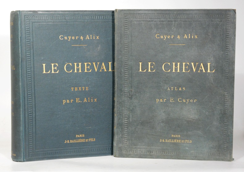
18860044451886 Paris, Baillière, 1886. Deux volumes grand in-quarto (230 X 290) percaline vert lierre, encadrements à froid sur les plats, auteur, titre et nom de l'éditeur dorés sur le premier plat et au dos, atlas contenant un portefeuille en toile verte et carton au second contreplat (reliure de l'éditeur). Volume de texte : XXIV-703 pages - Atlas : 48 pages, 16 planches en couleurs sur vélin fort et 8 accessoires. Cartonnage de l'atlas un peu défraichi.
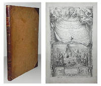
1685840Utrecht, Meinardus à Dreunen & Guilielmus à Walcheren, 1685. Mit gestoch. Titel, prachtvollem gefalt. Kupferstich-Porträt von J. Ebeling nach Romeyn de Hooghe und 16 gefalt. Kupfertafeln. 8 Bll (incl. gestoch. Titel), 568 Seiten, 3 Bll. Index; 8 Bll., 303 Seiten; 1 Bl., 130 Seiten, 1 Bl; 2 Bll., 70 Seiten. Folio. Lederband der Zeit mit reicher Rückenvergoldung.
In-4 p. (mm. 300x215), 3 volumi, brossura orig., sovraccoperta (con ingialliture, un dorso restaur. per mancanza), pp. XII,393; VIII,735c; VIII,1257 (numeraz. continua), con ritratto dell’autore e complessive 51 tavole, ciasc. con più figure, litografate in b.n. e in tinta (per lo più a doppia pag.), tutte dettagliatamente descritte. 1° vol. ”Istologia normale” (1870-1883) - 2° vol. “Istologia normale” (1883-1902) - 3° vol. “Patologia generale e isto-patologia” (1868-1894). Così è stata divisa in volumi la straordinaria raccolta dei lavori del sommo biologo Camillo Golgi, sempre sparsi in memorie pubblicate, a diverse epoche, in molti periodici ed atti di società scientifiche. "Prima edizione", in tiratura di 325 esemplari. Opera stampata, completa in 3 volumi, nel 1903 cui fece seguito, nel 1929, un 4° volume pubblicato postumo sempre da Hoepli, con il titolo "Scritti su argomenti vari" (1902-1925), a cura di L. Sala - E. Veratti - G. Sala. Cfr. Castiglioni “Storia della medicina”, pp. 784, 786, 798, 897: ”Camillo Golgi (Corteno, nel Bresciano, 7 luglio 1843 - Pavia, 21 gennaio 1926) fu, nel campo dell’anatomia del sistema nervoso, il più grande maestro del secolo XIX. Studiò medicina presso l’Università di Pavia, uno dei più antichi e prestigiosi atenei italiani, sotto la guida di Eusebio Oehl, grande studioso di anatomia ed istologia microscopica: rircercatore infaticabile, lavoratore diligentissimo, la sua fama attirava all’istituto dell’Università di Pavia allievi da ogni parte del mondo. Egli appartiene alla piccola schiera di coloro che lasciarono una traccia incancellabile nella storia della medicina dei nostri tempi.. Allievo di Giulio Bizzozero, iniziò nel 1870 i suoi studi sul tessuto di sostegno del sistema nervoso, ai quali seguirono una serie di altri studi come le ricerche sulla sostanza grigia cerebrale (1873) e sulla fine anatomia del cervello (1874); egli fu il primo ad indicare la colorazione cromo-argentea che rese possibili le indagini istologiche che condussero alla conoscenza della fine struttura dell’encefalo e del midollo spinale.. L’anatomia patologica del sistema nervoso centrale entrò nella sua fase moderna cogli studi del Golgi sulle alterazioni istologiche del cervello nella corea gesticolatoria e nell’idrofobia.. Un premio scientifico di alta importanza e di carattere internazionale, il premio Nobel, venne aggiudicato per la medicina a Golgi e Ramòn y Cajal nel 1906”. Cfr. anche Garrison and Morton che cita numerose opere del Golgi, fra cui, n. 1416: ‘Sulla fina anatomia degli organi centrali del sistema nervoso’ (Hoepli, 1886): “Golgi was en eminent Italian histologist. He demonstrated the existence of multipolar nerve-cells (Golgi cells) by means of his silver nitrate stain, and described the ‘Golgi appartus’ and ‘Golgi type II’ nerve cells - cells with short axons ramified within the cortex” e - n. 5239: ‘Sull’infezione malarica’: “Description of the development of the parasite of quartan malaria. Golgi differentiated the tertian and quartan parasites by the periods of their respective developments” - Bibliotheca Walleriana, n. 3629a - Dizionario Biograf. degli Italiani,LVII, pp. 599/613. Firma di apparten. sulle copertine e ai frontesp. dei 3 voll., con solo qualche lieve fiorit., altrimenti fresco esemplare, ben conservato.
173021397Venetiis apud Ballionem, 1730-1735 ; 4 tomes reliés en 6 volumes in-4° ; portrait gravé de Hoffman par J. Rupert aux tome I, IV (1) et tome I de “Consultationum” ; le tome 4 comprend trois parties soit les tomes IV, V et VI . [Suivi de] - Consultationum et responsorum medicinalium centuria prima complectens Morbos capitis et pectoris. 1 vol., 472 pp. - Consultatiomum et responsorum medicinalium centuria secunda et tertia complectens Morbos addominis et arturum externorum. 1 vol., 864, [54] pp. Francofurti ad Moenum, Franciscum Varremtrapp, 1734-1735; soit en tout 8 volumes in-4°, veau brun marbré, dos à nerfs très décorés et dorés, pièce de titre grenat, roulette dorée sur les coupes, tranches rouges ; les 2 derniers volumes sont légèrement plus grand et le décor du dos très légèrement différent.
174830143DBGöttingen, Abraham Vandenhoeck, 1748. 4°. (4) Bl., VI S., 136 S., (1) Bl. Mit 3 gestochenen, gefalteten Tafeln. Halbleinwandband um 1850 mit goldgeprägtem Rückentitel und Rückenvergoldung. + Wichtig: Für unsere Kunden in der EU erfolgt der Versand alle 14 Tage verzollt ab Deutschland / Postbank-Konto in Deutschland vorhanden +, A|B [2 Warenabbildungen]
In folio (mm. 495x329), mz. pelle coeva, decorazioni e tit. oro al dorso (restaur.), pp. VI,181,(5), con una serie di 22 straordinarie tavole anatomico-chirurgiche, a doppia pag., disegnate e inc. in rame da F. Anderloni: 11 al tratto e 11 in chiaroscuro. Vi sono contenute 5 Memorie: “Sull’ernia inguinale e scrotale - sulle sue complicazioni - sull’ernia femorale - sull’ernia gangrenata e sui mezzi che natura impiega per ristabilire la continuità del tubo intestinale - dell’ernia ombelicale e di quella della linea bianca dell’addome”. "Edizione seconda", accresciuta dall’autore di molte osservazioni anatomiche e patologiche. Cfr. Graesse,VI,291 - Brunet,V,183 - Premuda,190-191 - Sallander, p. 377 - Castiglioni, p. 614-16: "Scarpa (1752-1832) fu anatomico e chirurgo di grandissimo valore. Assieme allo Spallanzani, al Volta, al Frank, fu uno dei più illustri insegnanti alla scuola di Pavia che raggiunse verso la fine del '700 l'apice del suo splendore.. Fu anche disegnatore accurato e preciso: notevole fu la sua predilezione per l'iconografia quale insostituibile commento ed integrazione del testo”. Fresco esemplare ben conservato.
1 29.9x19.7 cm., [36], 340 pp., bel frontespizio inciso entro ricca cornice architettonica con figure di scheletri e vignette ai bordi e alla base, marca tipografica entro ovale, ripetuta in fine, ritratto dell'Autore, 46 bellissime tavv. anatomiche incise in rame n.t., legatura coeva in pergamena, titolo manoscritto al dorso, firme di appartenenza al frontespizio e in fine, lievissimi aloni ad alcune pagine, aloni pi? accentuati alla legatura, sporadiche fioriture, esemplare molto ben conservato, in latino Seconda edizione latina dopo quella del 1589. Valverde, medico spagnolo, studi? anatomia all'Universit? di Padova sotto Vesalio e Realdo Colombo. La sua Historia de la Composicion del Cuerpo Humano fu pubblicata per la prima volta a Roma nel 1556 anzich? in Spagna, tredici anni dopo l'apparizione della Fabrica di Vesalio. Valverde ha affermato nel suo testo di aver copiato le illustrazioni di Vesalio, ma quindici tavole sono opera sua, inoltre cambiato un certo numero di immagini del Vesalio combinandole o aggiungendo dettagli estranei come armature o nuove teste. Choulant-Frank attribuisce al pittore e scultore spagnolo Gaspar Becerra (1520-1570) le illustrazioni del libro di Valverde. Alcuni, come lo scorticato in piedi che tiene la propria pelle, o le porzioni di cadaveri sezionati che indossano armature, sono tra le immagini anatomiche pi? drammatiche del XVI secolo. Choulant-Frank pp. 207-08.
174830143DBGöttingen, Abraham Vandenhoeck, 1748. 4°. (4) Bl., VI S., 136 S., (1) Bl. Mit 3 gestochenen, gefalteten Tafeln. Halbleinwandband um 1850 mit goldgeprägtem Rückentitel und Rückenvergoldung.
174830143BBGöttingen, Abraham Vandenhoeck, 1748. 4°. 4 n.n. Bl., VI S., 136 S., 1 n.n. Bl. Mit 3 gestochenen, gefalteten Tafeln. Halbleinwandband um 1850 mit goldgeprägtem Rückentitel und Rückenvergoldung.